


Introduction
Congenital pseudarthrosis of the tibia (CPT) is one of the most biologically refractory conditions in pediatric orthopedics, affecting approximately 1 in 140,000–190,000 live births [1]. In neurofibromatosis type 1 (NF1)-associated cases – which account for 40–80% of all CPT – three pathological pathways converge simultaneously: Transforming growth factor-beta (TGF-β1)-driven fibrogenesis (“racing fibrosis”) via Smad2/3 and RhoA-MRTF-SRF; RAS-MEK-ERK hyperactivation secondary to NF1 biallelic loss in skeletal stem/progenitor cells (SSPCs), impairing osteoblastogenesis and elevating RANKL/OPG; and Wnt/β-catenin blockade via osteocyte-derived sclerostin overexpression [2,3]. Standard surgical fixation with an intramedullary rod or Ilizarov external fixation achieves primary union in 67–84% of cases, but refracture rates remain 22–48%, underscoring the persistent biological unmet need [4].
Umbilical cord mesenchymal stem cells (UC-MSC) secretome – comprising conditioned medium (CM), extracellular vesicles, and soluble factors – theoretically engages all three pathways simultaneously: anti-fibrotic via hepatocyte growth factor (HGF), prostaglandin E2 (PGE2), and exosomal miR-21-5p (TGF-βR suppression); pro-osteogenic via Wnt ligands, insulin-like growth factor 1, and bone morphogenetic protein (BMP-2/7) (Wnt/β-catenin rescue); and immunomodulatory via M1→M2 macrophage polarization [5,6]. Kurniawan et al. reported the only published clinical series of UC-MSC + secretome in CPT (n = 6; primary union 83%; final union 100% at 29 months) [7]. No published case has combined secretome with recombinant human BMP-2 (rhBMP-2), allogeneic UC-MSC whole cells, and a synthetic HA scaffold simultaneously, nor described a two-stage percutaneous secretome booster protocol.
We report the first case of this quadruple biological augmentation strategy with two-stage secretome delivery in NF1-confirmed Crawford type IV CPT of the distal tibia and concurrent fibular pseudarthrosis.
Case Report
Patient demographics and pre-operative assessment
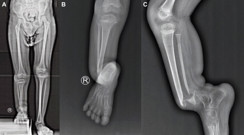
A 6-year-old male (age at surgery: 6 years 2 months) presented with a right distal tibial CPT and concurrent distal fibular pseudarthrosis (Crawford type IV), with no prior surgical history. Clinical examination revealed severe anterior bowing of the right distal tibia, inability to bear full weight independently, and restricted ankle range of motion secondary to the angular deformity. Limb length discrepancy was evident on standing anteroposterior (AP) whole-leg radiograph (Fig. 1a). Neurovascular status was intact distally. NF1 status was confirmed histopathologically from the hamartoma excision specimen: spindle cells with thin wavy nuclei, bland chromatin, and growth pushing host bone – consistent with neurofibromatosis, absent of atypia or anaplastic changes. Thoracolumbar radiographs showed no dystrophic scoliosis.
Pre-operative radiographs (Fig. 1b and c) demonstrated Crawford type IV CPT: Atrophic pseudarthrosis of the distal tibia with severe anterior bowing, sclerotic tapered bone ends, and concurrent pseudarthrosis of the distal fibula. The dual pseudarthrosis configuration eliminated the natural splinting role of an intact fibula, representing a particularly challenging biological and biomechanical environment. From a prognostic standpoint, NF1-associated Crawford type IV CPT with concurrent fibular pseudarthrosis carries the highest reported risk of refracture and non-union among all CPT subtypes [4,8].
Surgical procedure (November 19, 2025)
Under general anesthesia (operative duration: 3 h 15 min; blood loss: 50 mL; no transfusion), the following steps were performed sequentially:
(1) Longitudinal incision over the distal tibia with exposure of both tibial and fibular pseudarthrosis sites. (2) Radical excision of hamartomatous fibrous tissue until healthy bleeding bone ends were confirmed at both sites; the specimen was sent for histopathology (result: consistent with NF, as above). (3) Cross-pinning with K-wires at the distal tibial pseudarthrosis site only – no internal fixation at the fibular site. (4) Biological augmentation in sequence: Bongros (synthetic hydroxyapatite; osteoconductive scaffold) + Novosys (rhBMP-2; SMAD1/5/8-driven osteoblast differentiation, partially counteracting sclerostin-mediated Wnt blockade) + allogeneic UC-MSC whole cells (20 million cells; multipotent osteoprogenitor, sustained paracrine source, avoids autologous quality limitations in NF1-CPT [9]) packed into the defect, followed by UC-MSC CM (secretome; first injection) delivered as the final biological layer. (5) Application of Ilizarov external fixator with trans-ankle configuration: two tibial rings with half-pins + foot ring with calcaneal pins, enabling progressive compression at the tibial pseudarthrosis site. Post-operative management included antibiotic prophylaxis per institutional guideline and calcium/Vitamin D supplementation.
The delivery sequence (graft first, UC-MSC second, and secretome last) was chosen to: (a) establish a structural and osteoinductive scaffold before paracrine signaling; and (b) allow the secretome to function as a biological sealant, maximizing its concentration at the graft-bone interface.
Post-operative follow-up
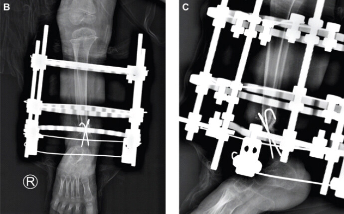
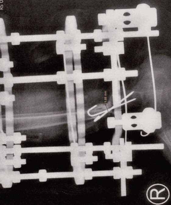
At 9 days, the patient was ambulatory with frame support (Fig. 2); pain had resolved (FLACC score 1, subsequently 0). At 6 months (May 16, 2026), radiological assessment demonstrated tibial callus formation with a measured AP diameter of 9.88 mm on lateral projection – equalling the width of the tibial diaphysis on the sagittal view – with no graft resorption (Fig. 3). The radiologist’s report confirmed no osteomyelitis and hardware in position; the distal tibiofibular pseudarthrosis was noted to show ongoing healing.
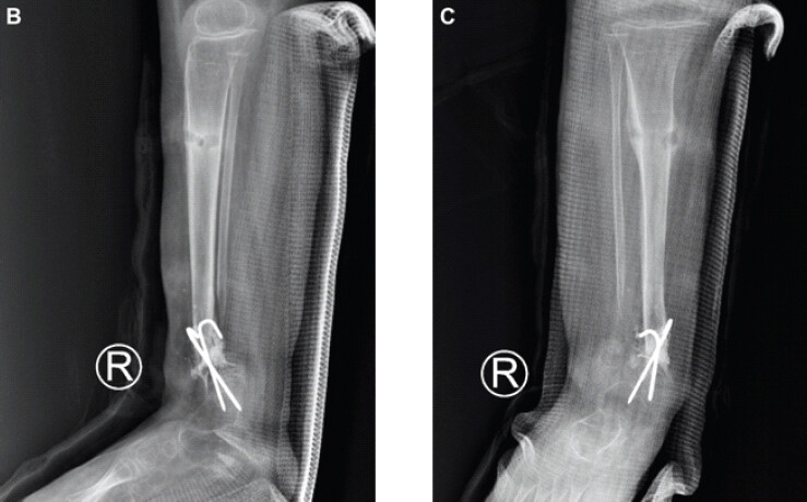
At 8 months (July 19, 2026), sufficient tibial bridging callus was confirmed clinically and radiologically to permit Ilizarov frame removal under general anesthesia (Fig. 4). At the time of removal, a second UC-MSC secretome injection (1.4 mL, percutaneous, fluoroscopy-guided) was delivered to the distal tibial union site to provide continued paracrine stimulation during the cortical remodeling phase. K-wires were retained at the distal tibial site. Post-removal radiographs demonstrated bridging callus at the tibial site with cortical continuity forming on both AP and lateral projections; anterior bowing was appreciably reduced; the fibular site showed ongoing healing without internal fixation or bridging. No refracture occurred. Pain score was 0 (NRS) at 8-month visit; the patient ambulated independently within the frame period. A patellofemoral-tibial brace was planned for post-removal rehabilitation. Summary of outcomes (Table 1).
Summary of clinical and radiological outcomes at 6 and 8 months postoperatively
| Outcome | Tibia | Fibula |
|---|---|---|
| 6-month callus | 9.88 mm AP diameter; no graft resorption | Not yet bridged |
| 8-month frame removal | Bridging callus; cortical continuity forming; alignment improved | Ongoing healing; no internal fixation |
| Refracture | None | None |
| Infection/hardware complication | None | None |
| Second secretome injection | 1.4 mL CM; percutaneous under fluoroscopy (Jul 19, 2026) | Not performed |
| Functional status | Frame-assisted ambulation; NRS 0 at 8 months; PTB brace planned | — |
PTB: Patellofemoral-tibial brace, AP: Anteroposterior, CM: Conditioned medium
Discussion
This case presents three interconnected novelties in managing NF1-associated CPT. First, it is the first reported application of quadruple biological augmentation – combining UC-MSC secretome, allogeneic UC-MSC, rhBMP-2, and a synthetic HA scaffold simultaneously – in CPT. Kurniawan et al. [7], the only prior clinical series of UC-MSC secretome in CPT, used UC-MSC + CM alone with locking plate fixation, achieving 83% primary union and 100% final union at a mean of 29 months (n = 6). The present protocol augments this approach with rhBMP-2 and a superior biomechanical construct (Ilizarov with dynamic compression), and adds a novel two-stage secretome delivery strategy.
Second, the two-stage secretome delivery – intraoperative (post-graft, as final biological layer) plus percutaneous fluoroscopy-guided booster at 8 months – has not been previously described in CPT. The rationale for the booster is mechanistically grounded: At 8 months, the maturing callus may benefit from renewed anti-fibrotic and pro-osteogenic paracrine stimulation to accelerate cortical remodeling and reduce the risk of delayed union at the tibial site.
Third, this is the first mechanistically annotated case systematically mapping each biological component to a specific CPT pathway. The secretome’s anti-fibrotic effect (TGF-βR suppression via HGF, PGE2, and exosomal miR-21-5p [5,6]) directly targets the racing fibrosis driven by NF1-deficient SSPCs in the residual periosteum – the mechanism that is not addressed by fixation, graft, or BMP-2 alone. The allogeneic UC-MSC source is preferred over autologous MSC in NF1-CPT patients: Dilogo et al. [9] demonstrated that autologous CPT-derived MSC quality is intrinsically compromised, likely secondary to NF1 haploinsufficiency in the marrow compartment. rhBMP-2 bypasses the sclerostin-mediated Wnt blockade via the SMAD1/5/8 axis [10], whereas Bongros provides the osteoconductive matrix through which both the secretome and BMP-2 signal. The Ilizarov frame provided dynamic compression at the tibial pseudarthrosis site, enabling piezoelectric and mechanotransduction stimulation of callus [4]; trans-ankle fixation immobilized the distal fibular site, compensating for the absence of natural fibular splinting in this dual-pseudarthrosis configuration.
The differential union response between tibia (bridging callus at 8 months) and fibula (ongoing healing) is attributable to multiple factors: (1) the tibial site received K-wire fixation providing mechanical stability, whereas the fibular site had no internal fixation; (2) a smaller fibular bone cross-section and lower BMP-2 delivery density at the fibular site; and (3) possible differential NF1 haploinsufficiency expression in fibular SSPCs. Future protocols should consider fibular-specific biological delivery.
Limitations include the single-case design, absence of a control arm, short follow-up (8 months insufficient to confirm durable union or assess long-term refracture risk), and undocumented secretome preparation parameters (CM volume, EV concentration, and passage number). These gaps are shared with Kurniawan et al. [7] and define the evidence requirements for future controlled trials. The mechanistic rationale for UC-MSC secretome in CPT triple-axis pathobiology has been reported by Kurniawan et al. [7].
Conclusion
Quadruple biological augmentation with UC-MSC secretome (two-stage delivery), allogeneic UC-MSC, rhBMP-2, and synthetic HA scaffold is clinically feasible in NF1-confirmed Crawford type IV CPT, with preliminary evidence of tibial bridging callus at 8 months without refracture. This protocol simultaneously targets the three convergent pathological pathways of NF1-associated CPT and introduces a novel two-stage secretome delivery strategy. Prospective controlled studies with standardized secretome dosimetry and longer follow-up are required to confirm efficacy.
Conflict of Interest:
Nil
Source of Support:
Nil
Consent:
The authors confirm that informed consent was obtained from the patient for publication of this article
How to Cite this Article
Anggitadewi LGD, Sindrawati O, Irianto KA, Martanto TW. Multimodal Biological Augmentation with Umbilical Cord Mesenchymal Stem Cell Secretome, Recombinant Human Bone Morphogenetic Protein-2, and Allogeneic Umbilical Cord Mesenchymal Stem Cells in Neurofibromatosis Type 1-Confirmed Congenital Pseudarthrosis of the Distal Tibia and Fibula: A Case Report with Mechanistic Scoping Review. Journal of Orthopaedic Case Reports 2026 October;16(10): 286-291.
References
- Pannier S. Congenital pseudarthrosis of the tibia. Orthop Traumatol Surg Res 2011;97:750-61. [Google Scholar] | [PubMed]
- Perrin S, Protic S, Bretegnier V, Radomska KJ, Laurendeau I, Colnot C. MEK-SHP2 inhibition prevents tibial pseudarthrosis caused by NF1 loss in Schwann cells and skeletal stem/progenitor cells. Sci Transl Med 2024;16:eadj1597. [Google Scholar] | [PubMed]
- Cho TJ, Seo JB, Lee HR, Yoo WJ, Chung CY, Choi IH. Biologic characteristics of fibrous hamartoma from congenital pseudarthrosis of the tibia associated with neurofibromatosis type 1. J Bone Joint Surg Am 2008;90:2735-44. [Google Scholar] | [PubMed]
- Kesireddy N, Kheireldin RK, Lu A, Cooper J, Liu J, Ebraheim NA. Current treatment of congenital pseudarthrosis of the tibia: A systematic review and meta-analysis. J Pediatr Orthop B 2018;27:541-50. [Google Scholar] | [PubMed]
- Liu C, Zhao Q, Zhu Q. Umbilical cord/placenta-derived mesenchymal stem cells inhibit fibrogenic activation in human intestinal myofibroblasts via inhibition of myocardin-related transcription factor A. Stem Cell Res Ther 2019;10:295. [Google Scholar] | [PubMed]
- Karner L, Jimenez W, Birkl-Toeglhofer AM. Anti-fibrogenic effect of umbilical cord-derived mesenchymal stem cell-conditioned media in human esophageal fibroblasts. Sci Rep 2024;14:22549. [Google Scholar] | [PubMed]
- Kurniawan A, Ivansyah MD, Dilogo IH, Hutami WD. Umbilical cord mesenchymal stem cells combined with secretome for treating congenital pseudarthrosis of the Tibia: A case series. Eur J Orthop Surg Traumatol 2023;33:2881-8. [Google Scholar] | [PubMed]
- Paley D. Congenital pseudarthrosis of the tibia: Biological and biomechanical considerations to achieve union and prevent refracture. J Child Orthop 2019;13:120-33. [Google Scholar] | [PubMed]
- Dilogo IH, Mujadid F, Nurhayati RW, Kurniawan A. Evaluation of bone marrow-derived mesenchymal stem cell quality from patients with congenital pseudoarthrosis of the tibia. J Orthop Surg Res 2018;13:266. [Google Scholar] | [PubMed]
- Das SP, Ganesh S, Pradhan S, Singh D, Mohanty RN. Effectiveness of recombinant human bone morphogenetic protein-7 in the management of congenital pseudoarthrosis of the tibia: A randomised controlled trial. Int Orthop 2014;38:1987-92. [Google Scholar] | [PubMed]
- Crawford AH, Schorry EK. Neurofibromatosis in children: The role of the orthopaedist. J Am Acad Orthop Surg 1999;7:217-30. [Google Scholar] | [PubMed]
- Giannoudis PV, Einhorn TA, Marsh D. Fracture healing: The diamond concept. Injury 2007;38 Suppl 4:S3-6. [Google Scholar] | [PubMed]
- Marsell R, Einhorn TA. The biology of fracture healing. Injury 2011;42:551-5. [Google Scholar] | [PubMed]
- Grill F, Bollini G, Dungl P, Fixsen J, Hefti F, Ippolito E. Treatment approaches for congenital pseudarthrosis of tibia: Results of the EPOS multicenter study. European Paediatric Orthopaedic Society (EPOS). J Pediatr Orthop B 2000;9:75-89. [Google Scholar] | [PubMed]
- Memeo A, Loiudice L, Stazzoni G. Treatment of congenital pseudoarthrosis of the tibia with bone marrow aspirate concentrate combined with external fixation. J Pediatr Orthop 2020;40 Suppl 1:S52-7. [Google Scholar] | [PubMed]
© 2026 Journal of Orthopaedic Case Reports - Published by Indian Orthopaedic Research Group
About the Authors
-

Luh Gede Djatu Anggitadewi
-

Oen Sindrawati
-

Komang Agung Irianto
-

Tri Wahyu Martanto
Related Articles in Journal of Orthopaedic Case Reports
 September 1, 2026 Angular Improvement with Bracing Over 7 Years in Anterolateral Tibial Bowing without Neurofibromatosis Type 1: A Case Report
 August 1, 2026 Oblique Plane Deformity Correction with Ilizarov Fixator in Ipsilateral Proximal Tibia Growth Arrest and Malunited Tibia: A Case Report
 February 1, 2026 Impaired Bone Healing and Fracture Complications during Limb Lengthening in a Neurofibromatosis Type 1 Patient Receiving Selumetinib: Case Report and Literature Review
 July 1, 2025 A Rare Case Report of Giant Cell Tumor in the Distal Tibia: Management with Tumor Excision and Limb Reconstruction with Ilizarov Fixator