

In internal fixation of patellar fractures, the orientation of the surgical incision meaningfully influences long-term cosmetic and functional recovery without altering fracture healing or complication risk.
Dr. Yash Agrawal, Department of Orthopaedics, Netaji Subhash Chandra Bose Medical College, Jabalpur, Madhya Pradesh, India. E-mail: yashagrawal876@gmail.com
Abstract
Introduction: Patellar fractures require internal fixation via a skin incision that must balance intraoperative exposure with cosmetic outcome. Vertical midline and horizontal curvilinear patella-centered incisions are both used, but comparative evidence on their functional and cosmetic outcomes remains limited.
Materials and Methods: A prospective, randomized comparative study was conducted on 60 patients undergoing internal fixation of patellar fractures, allocated equally to vertical midline (n = 30) or horizontal curvilinear (n = 30) incision groups by serial-number randomization. Patients were followed at 3, 6, 12, and 24 weeks, with functional outcome assessed by Oxford Knee Score, knee flexion, extension lag, and kneeling ability/comfort, and cosmetic outcome assessed by scar width, length, tenderness, pigmentation, and scar type. Data were analyzed using independent t-test and Chi-square test, with P < 0.05 considered significant.
Results: Baseline characteristics, intraoperative parameters, radiological union time, and complication rates were comparable between groups (P > 0.05). The vertical group showed transiently greater knee flexion at 6 weeks (P = 0.004), which equalized by 12–24 weeks. The horizontal group demonstrated significantly better outcomes at 24 weeks, including narrower and shorter scars, less tenderness, higher Oxford Knee Score (43.20 ± 2.46 vs. 36.13 ± 3.62; P < 0.001), and superior kneeling duration and comfort (P < 0.05). Normal scar healing at 24 weeks was more frequent with horizontal incision (80.0% vs. 46.7%), though not statistically significant (P = 0.124).
Conclusion: Horizontal curvilinear incision achieves functional recovery at least equivalent to, and cosmetic outcome superior to, vertical midline incision in patellar fracture fixation, without compromising surgical feasibility or safety.
Keywords: Patellar fracture, internal fixation, surgical incision, Oxford Knee Score, scar outcome, cosmesis.

Patellar fractures account for approximately 1% of all skeletal fractures and disproportionately affect older women and young active men, with population-based data showing incidence rates that rise steadily with advancing age [1]. Fracture morphology exhibits considerable variability, and recent multicenter fracture-mapping studies have demonstrated that comminuted and displaced patterns – the fracture types most commonly requiring open reduction and internal fixation (ORIF) – are prevalent across all age groups, underscoring the continued importance of surgical fixation in contemporary trauma practice. [1]. Restoring articular congruity and extensor mechanism continuity remains the primary surgical goal, and contemporary reviews of patella fracture management describe a wide range of fixation constructs and surgical exposures aimed at balancing secure fixation with minimization of soft-tissue morbidity [2]. Regardless of implant choice, however, adequate surgical exposure of the patella continues to depend on the skin incision, and functional recovery following ORIF is now benchmarked using validated patient-reported measures; recent series report mean post-operative Oxford Knee Scores in the mid-30s among patients treated for patellar fractures, underscoring that functional recovery, while generally favorable, still varies meaningfully across techniques and exposures [3].
Beyond fracture union and joint function, the cosmetic outcome of the surgical scar has emerged as an increasingly important determinant of patient satisfaction, particularly over a joint subjected to repetitive flexion-related tension. The direction in which a skin incision is placed relative to the natural tension lines of the skin is a recognized determinant of scar quality: incisions that run parallel or oblique to relaxed Langer’s lines are subjected to less mechanical stretch during healing and are less prone to hypertrophic widening, whereas incisions crossing these lines perpendicularly experience sustained tension that predisposes to hypertrophic and keloid scarring [4]. Notably, the knee has been specifically highlighted as a site where the anatomically ideal, low-tension incision line runs horizontally, yet vertical incisions are frequently used instead because they permit a wider surgical field, creating an inherent trade-off between exposure and cosmesis [4]. This principle has been empirically demonstrated in other areas of orthopedic surgery: A recent comparative study of total hip arthroplasty incisions found that transverse incisions aligned with relaxed skin tension lines produced significantly better patient-reported scar satisfaction and appearance scores than conventional longitudinal incisions, without compromising sensory outcomes [5].
Despite this evolving evidence in adjacent orthopedic procedures, comparative data specifically addressing incision orientation – vertical midline versus horizontal curvilinear, patella-centered – in the surgical management of patellar fractures remain limited, and existing literature on patellar fracture surgery has focused predominantly on fixation technique and union rates rather than on the influence of incision geometry on combined functional and cosmetic outcomes [1,2]. Given the anatomical and biomechanical similarities between the knee and hip in this regard, and the growing recognition that patient-reported cosmetic satisfaction is a legitimate surgical outcome in its own right, a direct comparison of vertical midline and horizontal curvilinear incisions in patellar fracture fixation is warranted [4,5,6]. This study was therefore undertaken to compare the functional and cosmetic outcomes of these two incision approaches in patients undergoing internal fixation of patellar fractures.
Ethical considerations
The study was conducted after approval from the Institutional Ethics Committee (No. IEC/2024/4539–111, dated May 16, 2024). Written informed consent was obtained from all participants; confidentiality was maintained throughout, and participation was voluntary, with patients free to withdraw at any stage without affecting their routine care.
Study design
This was a prospective, randomized, comparative study designed to evaluate and compare the functional and cosmetic outcomes of two surgical skin incisions – vertical midline and horizontal curvilinear patella-centered incisions – used during internal fixation of patellar fractures. Eligible patients were enrolled consecutively and allocated to one of two groups by a simple randomization method based on serial numbers. The primary focus was on post-operative functional recovery, scar healing and cosmetic outcome, and intraoperative technical feasibility (ease of reduction, implantation, and repair of the extensor mechanism).
Study setting
The study was conducted in the Department of Orthopaedics, N.S.C.B. Medical College and Hospital, and the Regional Spine Injury Center, Jabalpur (Madhya Pradesh, India) – tertiary referral center for trauma cases from both urban and rural regions of the state.
Study duration
The study was conducted over 2 years, allowing sufficient time for patient recruitment, surgical intervention, and post-operative follow-up up to 24 weeks for assessment of functional recovery, fracture union, and scar-related outcomes.
Inclusion criteria
- Patients aged 20–60 years admitted with patellar fracture
- Patients willing to give informed consent for participation in the study.
Exclusion criteria
- Patients unwilling to undergo surgery
- Patients with undisplaced fractures
- Patients with open (grade III) patellar fractures
- Patients with patellar fracture associated with internal derangement of the knee
- Patients with associated fractures of the ipsilateral femur, tibia, or other lower-limb injuries.
Only patients meeting the inclusion criteria and providing written informed consent were enrolled.
Sample size
The sample size was calculated using the standard formula for comparison of two independent means: n = 2σ2 (Zα/2 + Zβ)2/d2 where Zα/2 = 1.96 (for a two-sided α of 0.05), Zβ = 0.84 (for 80% power), σ = assumed pooled standard deviation of 6.5 points in Oxford Knee Score [3], and d = expected minimum clinically meaningful mean difference of 5 points between groups.
This yielded a calculated sample size of approximately 27 patients per group. Accounting for an anticipated 10% loss to follow-up, the final sample size was rounded up to 30 patients per group, for a total of 60 patients (Group A: horizontal curvilinear incision, n = 30; Group B: vertical midline incision, n = 30).
Study groups and randomization
Patients were randomized into two groups using a serial-number method. Both groups underwent standard internal fixation techniques appropriate to the fracture pattern.
Study parameters
The following parameters were evaluated:
- Pre-operative knee joint mobility
- Post-operative functional outcome (range of motion, Oxford Knee Score)
- Cosmetic outcome of the surgical scar (width, length, tenderness, pigmentation, keloid formation, patient satisfaction)
- Healing of the incision and complications (infection, dehiscence, hypertrophic scarring)
- Intraoperative ease of reduction, implantation, and extensor mechanism repair
- Radiological union, assessed by serial X-rays and ultrasonography.
Study procedure
On admission, all patients underwent clinical evaluation and hemodynamic stabilization, with routine laboratory investigations and radiological assessment (anteroposterior, lateral, and skyline knee radiographs). Patients were operated upon as soon as medically fit for surgery and anesthesia. All procedures were performed in the supine position under image intensifier guidance, using standard internal fixation techniques appropriate to fracture configuration. The assigned incision (vertical or horizontal) was used according to randomization, and intraoperative ease of exposure, reduction, implant placement, and extensor mechanism repair was recorded.
Postoperatively, patients received routine antibiotics and anti-inflammatory medication. A check radiograph was obtained on the 3rd post-operative day, sterile dressings were performed on the 3rd and 5th post-operative days, and sutures were removed between post-operative days 10–15, after which patients were discharged with standard rehabilitation advice.
Data collection
Data were collected using a structured pro forma. Baseline demographic and clinical details and pre-operative knee mobility were recorded at admission. Post-operative assessments were performed at 2, 6, 12, and 24 weeks (Figs. 1, 2, 3, 4), comprising clinical examination for pain, tenderness, scar condition, and range of motion; radiographic evaluation for fracture union; ultrasonography to assess adhesions or fluid collection; functional assessment using the Oxford Knee Score; and scar evaluation for width, length, tenderness, pigmentation, keloid formation, and discomfort on stretching.
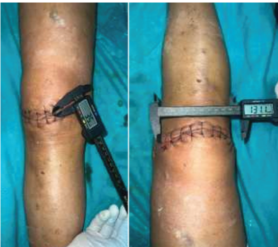
Figure 1: Horizontal incision (at 2 weeks).
Statistical analysis
All data were entered into a master chart and analyzed using the Statistical Package for the Social Sciences for Windows version 25.0. Quantitative variables were expressed as mean ± standard deviation and compared using the independent samples t-test. Qualitative variables were expressed as frequencies and percentages and compared using the Chi-square test. A P < 0.05 was considered statistically significant.
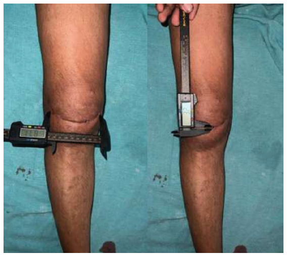
Figure 2: Horizontal incision (at 12 weeks).
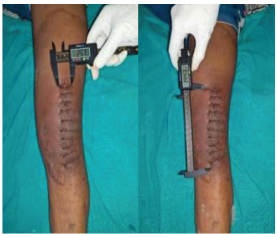
Figure 3: Vertical incision (at 2 weeks).
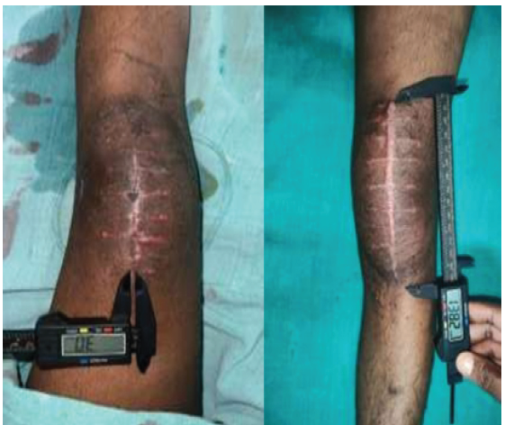
Figure 4: Vertical incision (at 12 weeks).
The two groups were comparable with respect to age distribution, sex, mode of injury, side of involvement, and fracture type, with no statistically significant differences observed for any baseline variable (all P > 0.05; Table 1), confirming adequate randomization.
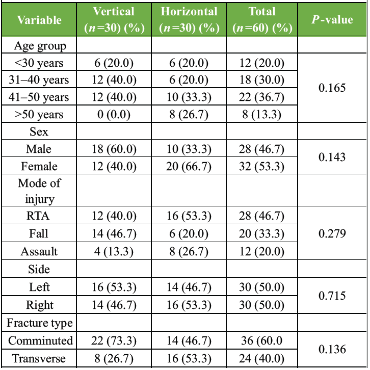
Table 1: Baseline demographic and clinical characteristics (n=30)
Duration of surgery (71.60 ± 10.50 vs. 72.00 ± 11.44 min), intraoperative blood loss (136.07 ± 32.39 vs. 138.60 ± 36.70 mL), and incision length (11.97 ± 1.45 vs. 12.21 ± 1.05 cm) were similar between the vertical and horizontal groups, respectively, with no statistically significant differences (P > 0.05 for all; Table 2), indicating comparable operative exposure and technical feasibility between the two approaches.
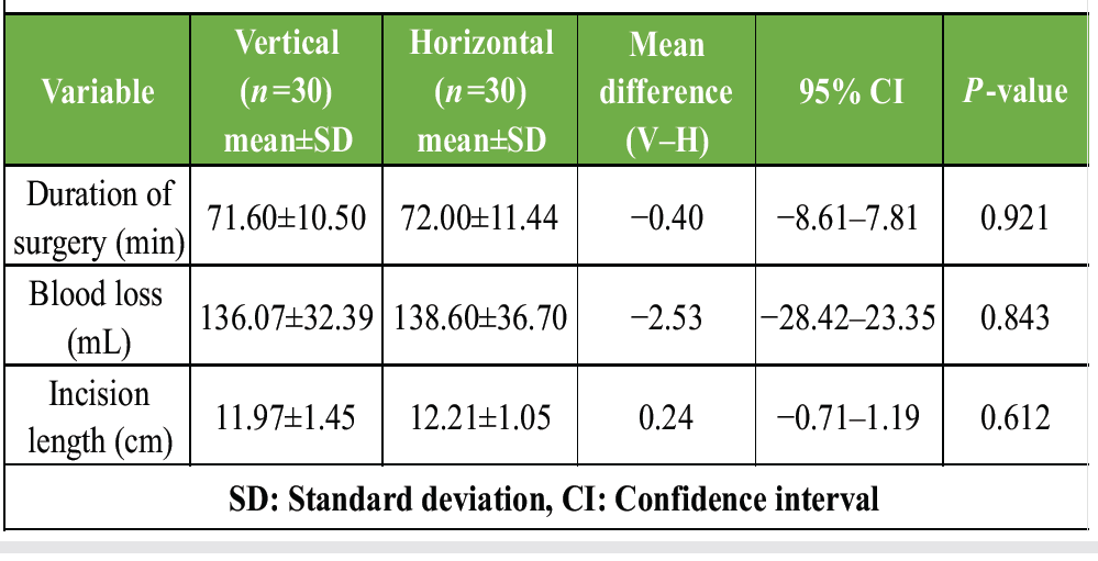
Table 2: Intraoperative parameters
Knee flexion was comparable between groups at 3 weeks (P = 0.874). At 6 weeks, the vertical incision group demonstrated significantly greater flexion than the horizontal group (87.73 ± 7.29 vs. 78.67 ± 8.56; P = 0.004). This difference resolved by 12 weeks (P = 0.386), and at 24 weeks the vertical group showed a non-significant trend toward higher flexion (128.53 ± 4.31 vs. 125.47 ± 3.91; P = 0.051). Extension lag improved progressively in both groups throughout follow-up, with no statistically significant between-group differences at any time point (P > 0.05; Table 3).
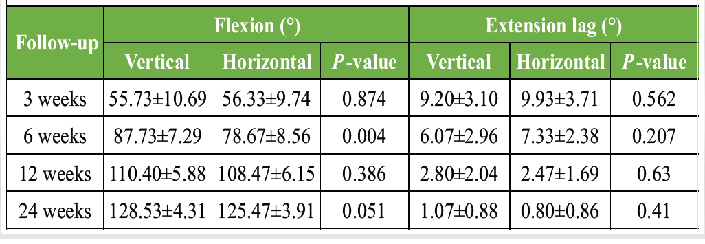
Table 3: Functional recovery – knee flexion and extension lag
Scar width and length were comparable between groups at 3, 6, and 12 weeks (P > 0.05). By 24 weeks, however, the horizontal incision group had significantly narrower scars (2.79 ± 0.62 mm vs. 4.30 ± 0.66 mm; P < 0.001) and shorter scars (10.00 ± 1.45 cm vs. 11.00 ± 1.05 cm; P = 0.045) compared to the vertical group. Scar tenderness (Visual Analog Scale) was significantly higher in the horizontal group at 3 weeks (5.53 ± 1.36 vs. 4.40 ± 1.12; P = 0.019) and again at 24 weeks (1.60 ± 0.74 vs. 0.67 ± 0.90; P = 0.004), with no significant difference at 6 or 12 weeks. Normal skin color (absence of hyperpigmentation) was significantly more frequent in the horizontal group at 3 weeks (80.0% vs. 33.3%; P < 0.001) and 6 weeks (73.3% vs. 33.3%; P = 0.028), with the difference narrowing and losing significance by 12 and 24 weeks (Table 4).
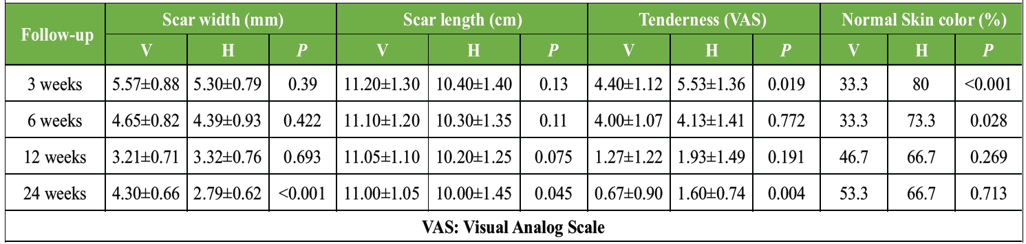
Table 4: Cosmetic and scar outcomes across follow-up
At 24 weeks, final scar type also favored the horizontal group: Normal scarring was seen in 80.0% of the horizontal group versus 46.7% of the vertical group, while keloid formation was more frequent in the vertical group (33.3% vs. 6.7%). This difference in overall scar type distribution did not reach statistical significance (χ² = 4.182, P = 0.124), likely reflecting the limited sample size, although the pattern was clinically consistent with the other cosmetic measures.
Oxford Knee Scores were similar between groups through 12 weeks (P > 0.05) but were significantly higher in the horizontal group at 24 weeks (43.20 ± 2.46 vs. 36.13 ± 3.62; P < 0.001). Kneeling ability (proportion able to kneel for ≥2 min) was significantly greater in the horizontal group at 3 weeks (53.3% vs. 26.7%; P < 0.001), 12 weeks (93.3% vs. 60.0%; P = 0.031), and 24 weeks (93.3% vs. 66.7%; P = 0.001). Kneeling comfort (proportion reporting high comfort) showed no significant difference at 3 or 6 weeks but was significantly higher in the horizontal group at 12 weeks (60.0% vs. 26.7%; P = 0.041) and 24 weeks (73.3% vs. 46.7%; P = 0.038) (Table 5).
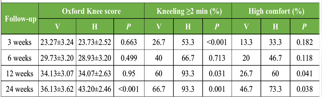
Table 5: Patient-centered functional outcomes across follow-up
Mean time to radiological union was comparable between the vertical and horizontal groups (12.87 ± 1.89 vs. 13.27 ± 2.52 weeks; P = 0.626). Overall post-operative complication rates were also similar (26.7% vs. 20.0%; χ2 = 0.186, P = 0.666), indicating that neither incision orientation increased operative morbidity (Table 6).
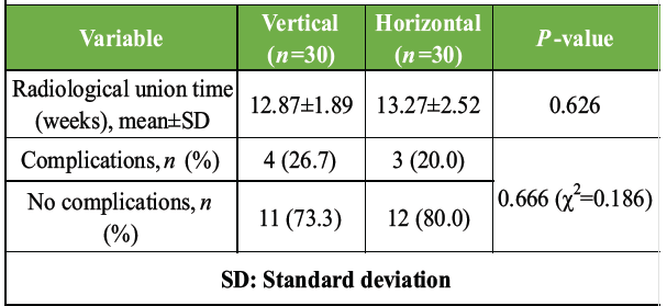
Table 6: Radiological union and post-operative complications
This prospective study compared vertical midline and horizontal curvilinear patella-centered incisions in patients undergoing internal fixation of patellar fractures and found that while early intraoperative and short-term functional parameters were comparable between the two approaches, the horizontal curvilinear incision was associated with significantly better cosmetic and patient-centered functional outcomes by 24 weeks (Tables 3, 4, 5, 6).
Baseline comparability between groups (Table 1) and equivalence of intraoperative parameters (Table 2) in our cohort mirror the pattern reported in other recent patellar fracture comparative series from India, where well-matched treatment arms are considered essential before attributing outcome differences to the surgical variable under study rather than to fracture severity or patient factors [7,8]. Similarly, our radiological union time of approximately 13 weeks in both groups (Table 6) closely parallels the union times of 12.75–12.85 weeks reported in a randomized Indian trial comparing tension band fixation materials for patellar fractures, reinforcing that incision orientation, unlike fixation construct, does not appear to influence the biological timeline of fracture healing [9].
The early advantage in knee flexion observed in the vertical incision group at 6 weeks (Table 3) is broadly consistent with reports that a vertical midline exposure offers more direct and generous access to the extensor mechanism, facilitating easier intraoperative repair and, in turn, earlier confidence in post-operative mobilization; this advantage of direct longitudinal exposure has also been highlighted in case reports of complex extensor mechanism repair, where a longitudinal incision was specifically chosen to allow adequate visualization of both fracture poles [10]. However, this early advantage was not sustained, and by 24 weeks, flexion was comparable between groups (Table 3) – a pattern also reported by Prasanna A et al. [7] in a South Indian series of comminuted patella fractures where excellent terminal flexion (mean 123°) and absence of extensor lag were achieved irrespective of the specific exposure used, suggesting that with adequate rehabilitation, ultimate range of motion converges regardless of the initial incision-related head start [7].
The most consistent and clinically meaningful finding in our study was the superiority of the horizontal curvilinear incision in cosmetic outcomes – narrower and shorter scars, less tenderness, and a favorable (though not statistically significant) shift toward normal scar healing rather than keloid formation at 24 weeks (Table 4). This is biologically plausible: Pathological scarring is now understood to be substantially driven by mechanical tension transmitted across the wound during healing, with high-tension anatomical sites and incisions that cross the predominant direction of skin stretch being disproportionately prone to hypertrophic and keloid change [11]. Because the knee is subjected to large, repetitive longitudinal skin tension during flexion, a vertical incision running parallel to this tension vector remains under sustained stretch throughout healing, whereas a horizontal curvilinear incision is comparatively unloaded – a mechanistic explanation consistent with the keloid clustering we observed in the vertical group (Table 4).
Our finding that the horizontal incision also conferred a significant functional advantage in Oxford Knee Score, kneeling duration, and kneeling comfort from 12 weeks onward (Table 5) is a more novel observation and stands in some contrast to a recent randomized controlled trial in total knee arthroplasty, which found that incision position (lateral versus midline) did not significantly affect kneeling ability or sensory outcomes at 6–12 months [12]. This discrepancy may reflect differences between an elective arthroplasty population and a trauma population with an acutely fractured, comminuted patella, where scar-related tenderness and hypertrophic change over the point of direct kneeling contact rather than sensory nerve disruption alone may be the dominant driver of kneeling difficulty, a mechanism not directly tested in the arthroplasty trial [13].
This study has several limitations. Although the sample size was prospectively calculated for the primary functional outcome, it was relatively small and therefore underpowered to detect differences in uncommon events such as infection, implant failure, or keloid formation. Furthermore, this was a single-center study conducted at a tertiary care institution, which may limit the generalizability of the findings to other healthcare settings, surgeons, and patient populations. The study also included only patients aged 20–60 years with displaced closed patellar fractures, limiting extrapolation of the results to elderly patients, open fractures, polytrauma, or fractures associated with ligamentous injuries.
The follow-up period of 24 weeks was sufficient to assess fracture union and early functional and cosmetic outcomes but may not fully capture long-term scar maturation, implant-related symptoms, functional recovery, or late complications. In addition, patients were allocated using a serial-number randomization method rather than a computer-generated randomization sequence with allocation concealment, introducing the potential for selection bias. Blinding of patients and outcome assessors was not feasible because the incision remained visible throughout follow-up, increasing the possibility of observer bias in subjective assessments. Cosmetic outcomes were evaluated using objective scar characteristics rather than validated scar assessment tools such as the Patient and Observer Scar Assessment Scale or Vancouver Scar Scale. Although a standardized rehabilitation protocol was advised, adherence, physiotherapy intensity, pain management, and patient compliance were not objectively monitored and may have influenced functional recovery. Finally, broader patient-reported outcomes, including health-related quality of life, return to work or sports, and long-term patient satisfaction, were not assessed. Future multicenter randomized studies with larger sample sizes, longer follow-up, validated outcome measures, and standardized rehabilitation monitoring are warranted to confirm these findings.
In patients undergoing internal fixation of patellar fractures, both vertical midline and horizontal curvilinear incisions provided comparable operative feasibility, with similar duration of surgery, blood loss, and radiological union time, and no difference in overall complication rates. While the vertical incision offered a modest early advantage in knee flexion at 6 weeks, this difference did not persist beyond 12 weeks. In contrast, the horizontal curvilinear incision was associated with significantly superior long-term outcomes at 24 weeks, including better Oxford Knee Scores, narrower and shorter scars, greater kneeling duration and comfort, and a clinically favorable, though not statistically significant, trend toward normal scar healing. These findings suggest that the horizontal curvilinear patella-centered incision achieves functional recovery at least equivalent to, and cosmetic outcomes superior to, the vertical midline approach, without compromising surgical feasibility or safety, supporting its consideration as a preferred incision choice in internal fixation of patellar fractures where cosmetic outcome is a priority.
In patients undergoing internal fixation of patellar fractures, the choice of incision carries meaningful clinical consequences beyond surgical exposure. Both vertical midline and horizontal curvilinear approaches are equally safe and feasible, with comparable operative time, blood loss, union, and complication rates. However, while the vertical incision offers only a brief early advantage in knee flexion, the horizontal curvilinear incision yields superior results by 12–24 weeks, with better functional scores, greater kneeling comfort, and a more favorable cosmetic profile. Surgeons managing patellar fractures – especially where cosmetic outcome matters – should consider the horizontal curvilinear incision as the preferred approach, without compromising fracture healing or safety.
References
- 1. Larsen P, Court-Brown CM, Vedel JO, Vistrup S, Elsoe R. Incidence and epidemiology of patellar fractures. Orthopedics 2016;39:e1154-8. [Google Scholar] [PubMed]
- 2. Bezerra CM, Bezerra CL. Current management of patellar fractures: Surgical innovations, conservative therapies, and rehabilitation strategies. Asclepius Int J Sci Health Sci 2025;4:293-312. [Google Scholar] [PubMed]
- 3. Feathers JR, Fellows D, Richardson E, Khatir M, George A, Ashwood N. Surgical outcomes following patella fracture repair: A single-center retrospective cohort study. Cureus 2025;17:e92343. [Google Scholar] [PubMed]
- 4. Huang C, Quong WL, Kamii Y, Ogawa R. Ideal surgical incision lines minimizing tension: A proposal based on observations of hypertrophic scars and keloids. Plast Reconstr Surg Glob Open 2025;13:e7344. [Google Scholar] [PubMed]
- 5. Kuramitsu Y, Itou J, Munakata Y, Okazaki K. Transverse incisions improve scar outcomes in anterolateral supine approach total hip arthroplasty: A patient observer scar assessment scale-based study. Arthroplasty 2026;8:2. [Google Scholar] [PubMed]
- 6. Yang S, Zhang E, Li Z, Long Y, Li Y, Zhang J, et al. Deep vein thrombosis in patients with patellar fractures: Assessing incidence rates and identifying risk factors. PLoS One 2025;20:e0316628. [Google Scholar] [PubMed]
- 7. Prasanna A, Arasu AD. Outcome analysis of fixed angle locking plate in comminuted patella fractures: A single centre prospective study from South India with early results. J Orthop Case Rep 2024;14:255-62. [Google Scholar] [PubMed]
- 8. Solunkhe S, Shah M, Patil V, Kuity K. Comparative study of inferior pole of patella fracture treated with transosseous technique using modified Krackow’s technique versus traditional anterior tension band wiring. J Orthop Case Rep 2024;14:230-5. [Google Scholar] [PubMed]
- 9. Noothan PT, Somashekara SA, Sunkappa SR, Karthik B, Rameshkrishnan K. A randomized comparative study of functional and radiological outcome of tension band wiring for patella fractures using SS wire versus fiberwire. Indian J Orthop 2023;57:876-83. [Google Scholar] [PubMed]
- 10. Mankar S, Jogewar R, Agrawal P. Non-union patella fracture – extensor mechanism repair with quadriceps release: A case report. J Orthop Case Rep 2024;14:55-9. [Google Scholar] [PubMed]
- 11. Fang X, Wang Y, Chen H, Yan Z, Jin S, Wu Y, et al. Hypertrophic scarring and keloids: Epidemiology, molecular pathogenesis, and therapeutic interventions. MedComm (2020) 2025;6:e70381. [Google Scholar] [PubMed]
- 12. Barroso Rosa S, Wilkinson M, McEwen P, Morse L, Grant A, Doma K, et al. Skin sensory alteration and kneeling ability following cruciate retaining total knee arthroplasty are not affected by the incision position: A randomised controlled trial of simultaneous bilateral surgery. J Exp Orthop 2023;10:145. [Google Scholar] [PubMed]
- 13. Qi H, Duan W, Jia R, Wang S, An M, Long Y. Risk factors for surgical site infection after patellar fracture surgery in the elderly. J Orthop Surg Res 2024;19:830. [Google Scholar] [PubMed]



Related Articles in Journal of Orthopaedic Case Reports
 October 1, 2026 Single-Stage Fixation of Bilateral Open Medial Hoffa Fractures Associated with Bilateral Patella Fracture and Bilateral Distal Radius Fractures: A Case Report
 October 1, 2026 Association between Coronal Mechanical Axis Correction and Early Functional Outcomes after Primary Total Knee Arthroplasty: A Retrospective Observational Study
 August 1, 2026 Clinical and Functional Outcomes of Posterior Cruciate Ligament Tibial Avulsion Fractures Treated with a Mini-Open Posterior Approach and Internal Fixation: A Case Series
 July 1, 2026 Isolated Humeral Trochlear Fracture without Associated Elbow Injury: A Rare Case Report