

Large segmental clavicle non-unions can be successfully treated with autologous iliac crest bone grafting and dual plating, restoring clavicular length and achieving durable union.
Dr. Joseph Salem-Hernández, Department of Orthopedic Surgery, Ponce Health Sciences University School of Medicine, PO Box 7004, Ponce, Puerto Rico 00717, USA. E-mail: joseph.salem@upr.edu
Abstract
Introduction: Clavicle fractures are common, and although most midshaft fractures heal successfully, large segmental defects are associated with an increased risk of nonunion. Clavicle nonunion is often severely disabling, with patients experiencing chronic pain, progressive deformity, reduced shoulder strength, and impaired upper-extremity function. The standard approach reserves non-vascularized iliac crest bone graft (ICBG) for defects up to about 3 cm, whereas larger defects are conventionally reconstructed with vascularized grafts. Reports of successful non-vascularized iliac crest grafting for very large segmental defects, particularly in the revision setting after prior failed fixation and grafting, are uncommon. We present this case to expand the documentation of non-vascularized reconstruction and to offer technical guidance for this challenging clinical scenario.
Case Report: A 40-year-old woman of Hispanic (Puerto Rican) background presented with right shoulder pain and functional limitation after two prior operations for a displaced midshaft clavicle fracture, which left a 4.5-cm segmental defect. She had a longstanding heavy smoking history. After confirmed smoking cessation and nutritional optimization, she underwent open reconstruction with a tricortical autologous iliac crest graft harvested using an ultrasonic bone scalpel, combined with orthogonal dual plating. At 2-year follow-up, she had full active range of motion, normal strength, restored clavicular length, and stable fixation, with no donor-site complications.
Conclusion: A large segmental midshaft clavicle defect exceeding the size usually considered suitable for non-vascularized grafting was reconstructed successfully with autologous ICBG and dual plating, with durable clinical and radiographic results. The case supports non-vascularized grafting as a reasonable option in selected patients, particularly where vascularized grafting is not readily available, and underscores the importance of addressing modifiable risk factors such as smoking before reconstruction. It adds to a small body of evidence and may broaden reconstructive options in orthopedic and trauma practice.
Keywords: Clavicle nonunion, large segmental bone defect, iliac crest bone graft, dual plating, case report.

Clavicle fractures are common injuries, accounting for approximately 5–12% of all adult fractures, with more than two-thirds involving the midshaft region [1]. Conventionally, these fractures were treated nonoperatively because of low reported nonunion rates of about 1% and concerns regarding surgical complications [2]. Recent evidence has challenged this approach, demonstrating higher nonunion rates and poorer functional outcomes in displaced midshaft fractures managed without surgery [3]. A meta-analysis reported nonunion in approximately 15% of nonoperatively treated patients compared with about 1% of those treated operatively [4]. Recognized risk factors for nonunion include smoking, comminution, advanced age, and displacement >2 cm [5].
Clavicle nonunion, although relatively uncommon, can be severely disabling, producing chronic pain, deformity, reduced shoulder strength, and impaired function, and such sequelae are common after clavicular injury [6,7]. In fact, the impact of clavicle nonunion on health-related quality of life may even exceed that of nonunion in long bones such as the tibia, femur, or humerus [8]. Large segmental defects causing shortening of more than 1.4–2 cm are more likely to be symptomatic and can impair abduction and flexion and reduce internal rotation [9,10].
The treatment of clavicle nonunion, particularly with segmental bone loss, often requires reconstructive procedures [6,11]. Although early approaches included resection as a salvage procedure, this is now avoided because of the risk of compromising shoulder girdle stability [6]. Modern surgical management involves open reduction and internal fixation (ORIF) using a range of implants, including intramedullary devices (e.g., Steinmann pins, Kirschner wires, and elastic nails), external fixators, and plates, particularly for displaced or complex injuries [6,12,13]. Although nonunion after ORIF is uncommon, when it occurs and becomes symptomatic, it can cause poor outcomes and functional impairment of the upper extremity [14].
Among reconstructive options, autologous bone grafting remains the gold standard, particularly using the iliac crest [15,16]. Iliac crest bone graft (ICBG) is commonly used to bridge defects of 2.5–3 cm; for defects larger than 3 cm and for cases in which previous surgery has failed, vascularized grafts are generally preferred [11]. Even so, ICBG is favored for its osteogenic, osteoconductive, and osteoinductive properties, which promote healing and integration [16]. Despite the potential for donor-site morbidity, major complications are rare [17], and harvesting from the iliac crest avoids the immunologic and infection-related risks associated with allografts and synthetic substitutes [18,19]. At present, there is no consensus on the management of large segmental defects exceeding 3 cm treated with ICBG.
Here, we present a rare case of recurrent clavicle nonunion with a 4.5-cm segmental defect successfully managed with ICBG. The case highlights the value of stable internal fixation with dual plating and the challenges of treating large bone defects after failed ORIF and bone grafting.
Initial presentation and previous surgeries
A 40-year-old woman presented to the orthopedic clinic with right shoulder pain, mild swelling, and tenderness on palpation over the mid-clavicle. The patient had undergone two previous procedures for a displaced midshaft clavicle fracture. The first was an ORIF with a plate construct, which failed because of poor fixation of the medial fragment. The second, performed 6 months before presentation to our clinic, involved removal of the fixation, debridement of the fracture site, and placement of demineralized bone matrix allograft in the defect. This sequence resulted in a 4.5-cm segmental defect of the mid-clavicle (Fig. 1).
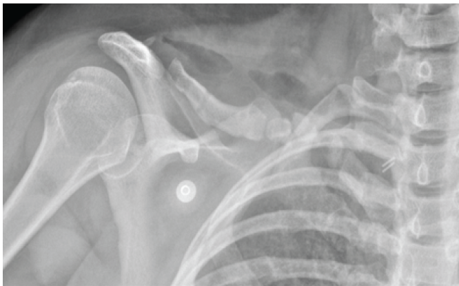
Figure 1: Pre-operative radiograph of the right clavicle showing a midshaft nonunion with a large segmental defect. Anteroposterior radiograph of the right clavicle obtained at initial presentation, demonstrating a midshaft nonunion with a large segmental bone defect of approximately 4.5 cm.
A detailed history and physical examination were performed, and new radiographs and computed tomography (CT) were obtained (Fig. 2).
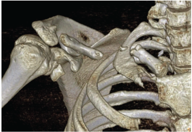
Figure 2: Three-dimensional computed tomography reconstruction of the right midshaft clavicle defect. Three-dimensional computed tomography reconstruction of the right clavicle at presentation, demonstrating the large segmental midshaft defect.
Passive range of motion (ROM) was preserved in all planes, but active motion was markedly limited and painful, with particular weakness in abduction and external rotation. She was unable to perform overhead activities or exercises such as bench pressing and front and side raises. The skin showed a well-healed transverse anterior clavicular incision with no fluid collection. Underlying infection was a concern, but serum inflammatory markers were negative, and the patient reported no soft-tissue healing problems after the previous procedures.
Smoking as a risk factor
The patient had a longstanding history of heavy tobacco use since the age of 14 years. Given the known association between smoking and impaired bone healing, she was counseled on smoking cessation before further surgery. After a 3-month cessation period confirmed by repeated negative nicotine testing, she was reassessed for definitive reconstruction. Calcium and Vitamin D supplementation was also prescribed.
Definitive surgery
The third procedure was an open repair of the clavicle nonunion with autologous ICBG and dual plating. The patient was positioned in the beach-chair position, and the upper right extremity and the contralateral iliac crest region were prepared and draped in sterile fashion. While the lead shoulder surgeon performed the clavicular reconstruction, a second surgeon harvested graft from the contralateral iliac crest. The defect was identified and thoroughly debrided to determine the final defect length, which was confirmed against measurements of the contralateral clavicle.
An ultrasonic bone scalpel (BoneScalpel; Misonix, Farmingdale, New York, USA) was used to make precise cuts in the iliac crest and to obtain a tricortical autograft of the required dimensions, measuring 4.5 cm in length and 1.8 cm in diameter, which fit well within the defect. Temporary Kirschner wire fixation (Fig. 3) was followed by spanning dual plating using two stainless-steel plates (Evos Mini Plating System; Smith and Nephew, Birmingham, Alabama, USA) applied in an anterior and superior (orthogonal) configuration (Fig. 4).
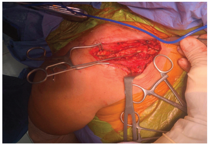
Figure 3: Intraoperative photograph of the iliac crest graft with temporary Kirschner wire fixation. Intraoperative photograph of the right clavicle during definitive reconstruction, showing the 4.5-cm tricortical autologous iliac crest bone graft within the segmental defect, held with temporary Kirschner wire fixation.
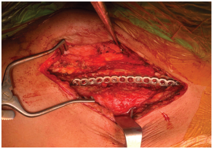
Figure 4: Intraoperative photographs of anterior and superior plate fixation. Intraoperative photographs during definitive reconstruction of the right clavicle. (A) Anterior plate spanning the medial native clavicle, the iliac crest graft, and the lateral clavicle. (B) Superior plate providing supplemental orthogonal fixation with additional screws.
Post-operative course and outcome
Immediate post-operative radiographs demonstrated dual-plate fixation with the interposed iliac crest graft (Fig. 5).
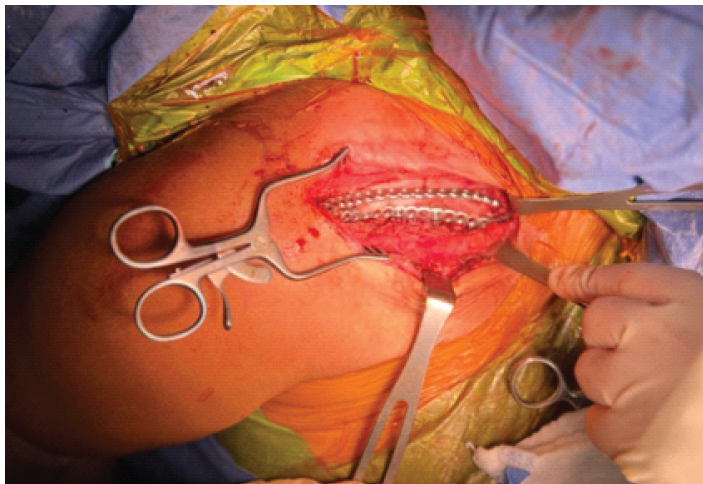
Figure 5: Immediate post-operative radiograph showing dual-plate fixation and grafting. Post-operative anteroposterior radiograph of the right clavicle, demonstrating dual-plate fixation of the midshaft nonunion with the autologous iliac crest bone graft.
The patient tolerated the post-operative period well and was placed in a sling for the first 6 weeks, allowing early passive ROM and Codman exercises. Physical therapy was prescribed, and active ROM was permitted at 6 weeks. Gentle strengthening was introduced at 2 months. At 6-month follow-up, she had regained full active ROM in all planes, although strength remained slightly reduced compared with the contralateral side, and additional strengthening therapy was prescribed. Radiographs at that time showed restored clavicular length comparable to the contralateral side and well-positioned implants. Graft incorporation was difficult to assess radiographically because of the dual-plate construct, and CT was not considered clinically necessary. At final follow-up 2 years postoperatively, the patient had full active ROM in all planes with strength equal to the contralateral side. Radiographs confirmed maintained clavicular length, alignment, and fixation without hardware migration, consistent with graft incorporation (Fig. 6).
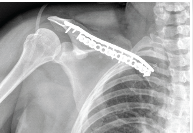
Figure 6: Two-year post-operative radiograph showing restored clavicular length and alignment. Anteroposterior radiograph of the right clavicle 2 years after reconstruction, demonstrating restored clavicular length and alignment compared with the contralateral side, with maintained fixation.
This case demonstrates successful reconstruction of a large segmental midshaft clavicle defect measuring 4.5 cm with autologous ICBG. The defect represented nearly one-third of the reported average female clavicle length of 15.1 cm [20]. Although ICBG is generally considered appropriate for defects of 2.5–3 cm, with larger defects typically reconstructed using vascularized grafts [11], this case supports the potential of ICBG as an effective alternative for larger defects.
Although ICBG is criticized for donor-site morbidity, including chronic pain, gait disturbance, hypertrophic or painful scarring, and nerve injury [21], our patient experienced no donor-site complications at 2 years. The graft was harvested with a high-frequency ultrasonic bone scalpel, which allowed precise harvest while minimizing adjacent soft-tissue trauma. This favorable outcome is consistent with a meta-analysis by Allsopp et al., which questioned the necessity of vascularization for longer bone grafts [22]. Vascularized grafts also carry potential disadvantages, including longer operative times, increased infection risk, and longer hospital stays [23]. In this context, ICBG may offer a simpler and safer alternative with favorable outcomes in selected patients, particularly in settings where vascularized grafting is not routinely performed.
With respect to fixation, dual plating has demonstrated biomechanical superiority over single plating, with reported improvements in union rates, reduced implant irritation, and a lower incidence of nonunion [14]. Advances in surgical technique have further contributed to improved functional recovery and fewer complications compared with non-operative treatment [10,24,25].
This case also illustrates several common risk factors for clavicle nonunion, including comminution, displacement >2 cm, and a long history of heavy smoking. Nonunion after ORIF for displaced midshaft fractures is generally uncommon but increases substantially in the presence of such risk factors [5,11]. The patient’s smoking history, large segmental defect, and prior hardware failure likely contributed to delay healing. Importantly, smoking cessation before and throughout the post-operative period may have supported improved healing.
This report has limitations. As a single case, the findings cannot be generalized, and studies with larger cohorts are needed to confirm the role of ICBG as an alternative to vascularized grafting for large segmental clavicle defects. A further limitation is that graft incorporation could not be confirmed with advanced imaging. Nevertheless, the case demonstrates that ICBG may be a viable option for large midshaft clavicle defects and supports further investigation.
This case demonstrates successful treatment of a recurrent midshaft clavicle nonunion with a large 4.5-cm segmental defect using autologous ICBG and dual-plate fixation. Despite a defect size exceeding the range traditionally considered suitable for non-vascularized grafting, the patient regained clavicular length and achieved complete functional recovery at 2-year follow-up. Attention to modifiable risk factors, particularly smoking cessation, together with stable dual-plate fixation, likely contributed to this favorable outcome. The case is of interest to orthopedic and trauma surgeons and suggests that autologous ICBG may broaden the reconstructive options available for large segmental clavicle defects, warranting further study.
Large segmental midshaft clavicle non-unions, even those exceeding 3 cm, can be reconstructed successfully with autologous tricortical iliac crest bone graft and orthogonal dual plating. Optimizing modifiable risk factors such as smoking and ensuring stable fixation are central to achieving durable union and functional recovery, and non-vascularized grafting may be a practical alternative to vascularized grafting in selected patients.
References
- 1. Postacchini F, Gumina S, De Santis P, Albo F. Epidemiology of clavicle fractures. J Shoulder Elbow Surg 2002;11:452-6. [Google Scholar] [PubMed]
- 2. Neer CS. Nonunion of the clavicle. JAMA 1960;172:1006-11. [Google Scholar] [PubMed]
- 3. Hill JM, McGuire MH, Crosby LA. Closed treatment of displaced middle-third fractures of the clavicle gives poor results. J Bone Joint Surg Br 1997;79:537-9. [Google Scholar] [PubMed]
- 4. McKee RC, Whelan DB, Schemitsch EH, McKee MD. Operative versus nonoperative care of displaced midshaft clavicular fractures: A meta-analysis of randomized clinical trials. J Bone Joint Surg Am 2012;94:675-84. [Google Scholar] [PubMed]
- 5. Murray IR, Foster CJ, Eros A, Robinson CM. Risk factors for nonunion after nonoperative treatment of displaced midshaft fractures of the clavicle. J Bone Joint Surg Am 2013;95:1153-8. [Google Scholar] [PubMed]
- 6. Martetschläger F, Gaskill TR, Millett PJ. Management of clavicle nonunion and malunion. J Shoulder Elbow Surg 2013;22:862-8. [Google Scholar] [PubMed]
- 7. Nowak J, Holgersson M, Larsson S. Sequelae from clavicular fractures are common. Acta Orthop 2005;76:496-502. [Google Scholar] [PubMed]
- 8. Schottel PC, O’Connor DP, Brinker MR. Time trade-off as a measure of health-related quality of life: Long bone nonunions have a devastating impact. J Bone Joint Surg Am 2015;97:1406-10. [Google Scholar] [PubMed]
- 9. Hillen RJ, Burger BJ, Pöll RG, De Gast A, Robinson CM. Malunion after midshaft clavicle fractures in adults: The current view on clavicular malunion in the literature. Acta Orthop 2010;81:273-9. [Google Scholar] [PubMed]
- 10. Patel B, Gustafson PA, Jastifer J. The effect of clavicle malunion on shoulder biomechanics: A computational study. Clin Biomech 2012;27:436-42. [Google Scholar] [PubMed]
- 11. Van der Burg FA, Baltes TP, Kloen P. Large segmental defects in midshaft clavicle nonunion treated with autologous tricortical iliac crest bone graft. Shoulder Elbow 2023;15:45-53. [Google Scholar] [PubMed]
- 12. Endrizzi DP, White RR, Babikian GM, Old AB. Nonunion of the clavicle treated with plate fixation: A review of forty-seven consecutive cases. J Shoulder Elbow Surg 2008;17:951-3. [Google Scholar] [PubMed]
- 13. Zlowodzki M, Zelle BA, Cole PA, Jeray K, McKee MD, Evidence-Based Orthopaedic Trauma Working Group. Treatment of acute midshaft clavicle fractures: Systematic review of 2144 fractures. J Orthop Trauma 2005;19:504-7. [Google Scholar] [PubMed]
- 14. Jenkins S, Daji A, Fomunung C, Grunhut J, Sabesan V. Managing recurrent clavicle nonunion and construct failure: A case report. J Orthop Exp Innov 2023;4:???. [Google Scholar] [PubMed]
- 15. Schmidt AH. Autologous bone graft: Is it still the gold standard? Injury 2021;52 Suppl 2:S18-22. [Google Scholar] [PubMed]
- 16. Campana V, Milano G, Pagano E, Barba M, Cicione C, Salonna G, et al. Bone substitutes in orthopaedic surgery: From basic science to clinical practice. J Mater Sci Mater Med 2014;25:2445-61. [Google Scholar] [PubMed]
- 17. Banwart JC, Asher MA, Hassanein RS. Iliac crest bone graft harvest donor site morbidity. A statistical evaluation. Spine 1995;20:1055-60. [Google Scholar] [PubMed]
- 18. Jones AL, Bucholz RW, Bosse MJ, Mirza SK, Lyon TR, Webb LX, et al. Recombinant human BMP-2 and allograft compared with autogenous bone graft for reconstruction of diaphyseal tibial fractures with cortical defects. A randomized, controlled trial. J Bone Joint Surg Am 2006;88:1431-41. [Google Scholar] [PubMed]
- 19. Szpalski M, Gunzburg R. Recombinant human bone morphogenetic protein-2: A novel osteoinductive alternative to autogenous bone graft? Acta Orthop Belg 2005;71:133-48. [Google Scholar] [PubMed]
- 20. Bernat A, Huysmans T, Van Glabbeek F, Sijbers J, Gielen J, Van Tongel A. The anatomy of the clavicle: A three-dimensional cadaveric study. Clin Anat 2014;27:712-23. [Google Scholar] [PubMed]
- 21. Boehm KS, Al-Taha M, Morzycki A, Samargandi OA, Al-Youha S, LeBlanc MR. Donor site morbidities of iliac crest bone graft in craniofacial surgery: A systematic review. Ann Plast Surg 2019;83:352-8. [Google Scholar] [PubMed]
- 22. Allsopp BJ, Hunter-Smith DJ, Rozen WM. Vascularized versus nonvascularized bone grafts: What is the evidence? Clin Orthop Relat Res 2016;474:1319-27. [Google Scholar] [PubMed]
- 23. Pogrel MA, Podlesh S, Anthony JP, Alexander J. A comparison of vascularized and nonvascularized bone grafts for reconstruction of mandibular continuity defects. J Oral Maxillofac Surg 1997;55:1200-6. [Google Scholar] [PubMed]
- 24. Von Rüden C, Rehme-Röhrl J, Augat P, Friederichs J, Hackl S, Stuby F, et al. Evidence on treatment of clavicle fractures. Injury 2023;54 Suppl 3:110818. [Google Scholar] [PubMed]
- 25. Xu J, Xu L, Xu W, Gu Y, Xu J. Operative versus nonoperative treatment in the management of midshaft clavicular fractures: A meta-analysis of randomized controlled trials. J Shoulder Elbow Surg 2013;23:173-81. [Google Scholar] [PubMed]




Related Articles in Journal of Orthopaedic Case Reports
 July 1, 2026 Three-column Plating for Complex Tibial Plateau Fractures: Multivariate Predictors of Superior Functional Recovery
 April 1, 2026 Modified Posterior Approach for Scapular Body Non-union: A Case Report
 April 1, 2026 Chondromyxoid Fibroma of the Right Thumb: Successful Management with Wide Local Excision, Iliac Crest Bone Grafting and K-Wiring – A Rare Case Report
 October 1, 2025 Surgical Treatment of a Non-union of Medial End Clavicle Fracture with Dual Plating and its Clinical Outcome – A Case Report