

Volar carpometacarpal fracture-dislocations are easily missed but can be successfully managed with acute closed reduction and percutaneous pinning to restore central column stability and achieve excellent early functional recovery.
Dr. Riad Fakih, Department of Orthopaedic Surgery, Clemenceau Medical Center, Dubai, United Arab Emirates. E-mail: riadrfakih@gmail.com
Abstract
Introduction: Volar dislocations of the carpometacarpal (CMC) joints are notably infrequent, attributable to the inherent stability afforded by their osseous morphology and robust volar ligamentous restrictions. When combined with fractures of two or more metacarpal bases, diagnosis and treatment may be delayed.
Case Report: A 43-year-old male patient presented with volar dislocation of multiple CMC joints and metacarpal fractures, involving the bases of the second, third, and fourth metacarpals, subsequent to a low-energy fall from a bicycle. The injury was evaluated utilizing plain radiographs and computed tomography scanning. The patient was successfully managed with closed reduction and percutaneous Kirschner wire fixation, achieving stable anatomical fixation without requiring open reduction. Post-operative imaging demonstrated well-preserved reduction and stable fixation, with no neurovascular complications. At 10 weeks postoperatively, the patient demonstrated near-symmetric range of motion and excellent clinical satisfaction.
Conclusion: CMC fracture dislocations are injuries that can be missed, and a high index of suspicion is mandatory in severe swelling beyond the radiological findings. Early identification and immediate stabilization of unstable volar CMC fracture-dislocation lead to successful therapeutic outcomes. Closed reduction and percutaneous fixation result in lower morbidity and better recovery by preserving soft tissue, re-establishing joint congruity, and restoring optimal hand kinematics.
Keywords: Volar carpometacarpal dislocation, metacarpal base fracture, closed reduction, percutaneous fixation, hand trauma.

Carpometacarpal (CMC) dislocations are infrequent injuries, comprising < 1% of hand trauma injuries [1,2]. The majority present dorsally, owing to the relative strength of the dorsal capsuloligamentous complex. Volar CMC dislocations are rare because of the strong volar ligaments, flexor tendon structures, and interlocking mechanics provided by the distal carpal row [3,4,5]. These fractures are commonly overlooked at the time of admission, as swelling, pain, and overlapping bony anatomy prevent appreciation of subtle radiographic findings. If untreated or inadequately managed, this condition may result in chronic instability, diminished grip strength, and wrist arthropathy [3,4]. This report presents an atypical volar CMC fracture-dislocation involving multiple metacarpal bases, along with its therapeutic approach.
An otherwise healthy 43-year-old male presented with pain and swelling in the left non-dominant wrist and hand after a low-velocity bicycle fall.
Clinical examination
Examination showed severe swelling, wrist deformity, and painful, limited motion, which was pronounced with passive ulnar and dorsal range of motion. Neurovascular examination was normal. There was no evidence of compartment syndrome of the left hand.
Imaging
Initial plain radiographs demonstrated a loss of normal CMC alignment with marked volar displacement and associated fractures at the bases of the second, third, and fourth metacarpals (Fig. 1).
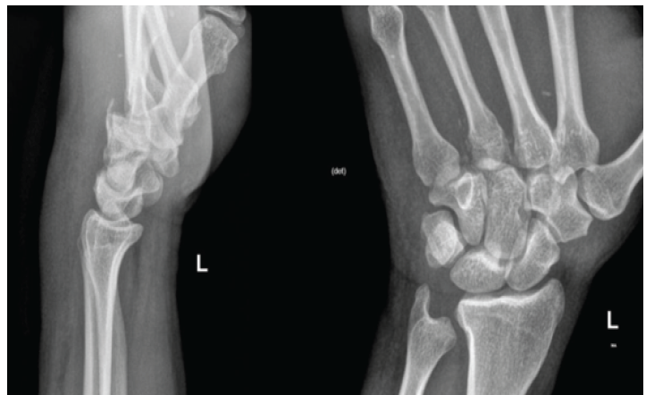
Figure 1: Pre-operative lateral and anteroposterior radiographs of the left hand demonstrating loss of normal carpometacarpal alignment. There is notable volar displacement of the metacarpal bases, accompanied by fractures at the bases of the second, third, and fourth metacarpals.
Given the complex injury pattern and the limited diagnostic sensitivity of plain radiographs for CMC dislocations, a computed tomography (CT) scan was performed. The scan demonstrated volar dislocation of the second, third, and fourth CMC joints, concomitant with dorsal intra-articular metacarpal base avulsion fractures. In addition, CT imaging is crucial in these injuries to rule out concurrent carpal bone fractures associated with metacarpal dislocations [6].
Surgical technique
Surgical stabilization of the fracture dislocation was performed. Under general anesthesia, the left upper extremity was scrubbed and draped, and a tourniquet was applied in case closed reduction fails. The dislocation was reduced after longitudinal traction and manipulation under fluoroscopic control. The fracture dislocation was evaluated for stability and was found to be unstable. Percutaneous fixation was achieved using several 2.0-mm Kirschner wires:
- From the second metacarpal into the capitate in a retrograde direction
- From the third metacarpal into the capitate, converging with the first K wire in a retrograde direction
- From the trapezium to the base of the second metacarpal in an antegrade direction.
This sequence of K-wire fixation has been shown to restore sagittal and rotational stability between the CMC joints [3,4,5]. Fluoroscopy confirmed anatomical reduction. A short arm cast was then applied, and post-operative anteroposterior and lateral radiographs demonstrated successful stabilization of the central column (Fig. 2).
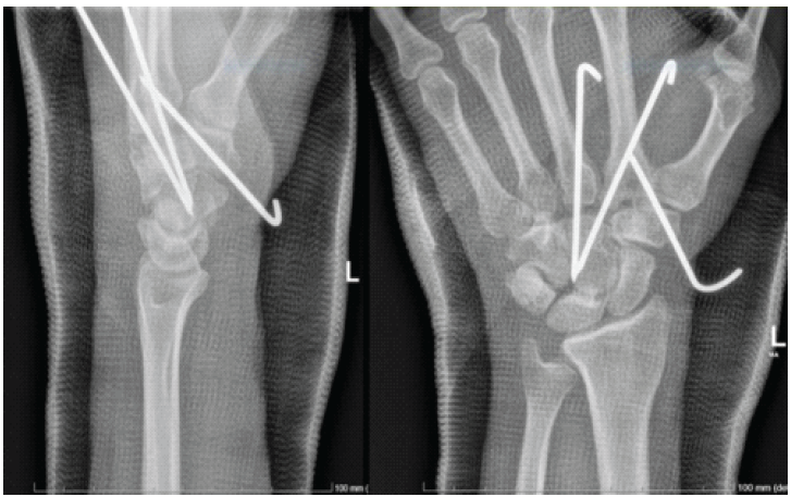
Figure 2: Post-operative anteroposterior and lateral radiographs demonstrating successful closed reduction and percutaneous fixation. Fixation was achieved using 2.0-mm Kirschner wires: bridging the second and third metacarpals to the capitate in a retrograde direction, and from the trapezium to the base of the second metacarpal in an antegrade direction.
Post-operative management
The patient was admitted overnight for clinical observation to monitor for compartment syndrome and cast-related compressive complications. Immobilization continued for 6 weeks, with weekly follow-up to assess swelling and finger range of motion. K-wires were removed after 6 weeks, and physical therapy was started immediately afterward. Follow-up radiographs obtained at 10 weeks postoperatively, 4 weeks following K-wire removal, revealed well-preserved reduction, maintained CMC alignment, and progressive interval healing (Fig. 3).
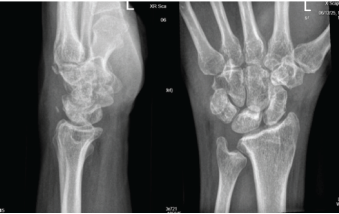
Figure 3: Follow-up anteroposterior and lateral radiographs at 10 weeks post-operative, following the removal of the Kirschner wires at the 6-week mark. The images demonstrate well-preserved reduction, maintained alignment of the carpometacarpal joints, and interval healing.
Early functional outcome
At the 10-week post-operative follow-up, the patient demonstrated nearly symmetric range of motion of the fingers, hand, and wrist compared to the uninjured side with limitation in ulnar deviation of the wrist and affected digits. The patient reported excellent functional recovery and return to work but was not yet cleared for return to sport. This was also reflected by excellent clinical satisfaction.
The CMC joints are stabilized by an interplay of osseous geometry, ligamentous restraint, and dynamic tendon forces [3,4,5]. The second and third CMC joints make up the stable, central column of the hand, supported by the trapezoid and capitate bones, whereas the fourth and fifth CMC joints compose a more mobile ulnar column that allows powerful grasping or cupping of the hand [3,4]. Dislocations through the volar CMC joint are uncommon because of the opposing volar ligaments and the flexor tendon forces on the palmar aspect, which tend to limit that direction of motion [3,4,5]. A combination of axial loading, wrist flexion, and rotational forces is usually necessary for volar displacement to occur [4,5,7]. In the current case, transmitted axial loading with a flexed wrist likely produced an excessive palmar shear force that overcame these restraints and caused failure in both the rigid central column and the mobile ulnar column. Concomitant fractures of metacarpal bases further destabilize the system by loss of buttressing from intact articular surfaces and increased susceptibility to displacement under physiologic forces [3]. Chronic dislocations change the loading on CMC joints with subsequent abnormal joint contact pressures and are at risk for chronic instability as well as post-traumatic arthritis [3,4,8]. Closed reduction can be performed acutely and is critical to regaining central joint alignment without jeopardizing surrounding soft tissue or the few remaining ligamentous attachments to maintain stability. This may be sufficient to neutralize shear and rotational forces during early healing when fixation bridges the central column using percutaneous K-wire fixation [4,5]. This procedure restores longitudinal stability with minimal surgical morbidity. Open reduction is required in chronic or irreducible cases, as it increases soft-tissue damage and stiffness [3,9].
While recent literature continues to emphasize that CMC fracture-dislocations typically result from high-energy mechanisms like motor vehicle collisions [10], our case demonstrates that these severe injuries can still occur following lower-energy bicycle falls. Furthermore, recent studies highlight that while open reduction is often required for delayed presentations or irreducible joint fragments, acute injuries that are anatomically reducible can be managed successfully with closed reduction and percutaneous pinning, yielding excellent functional recovery [11].
In the case of acute reducible injuries, closed reduction and percutaneous fixation offer a biomechanically viable and less invasive option. This approach minimizes soft tissue dissection and stiffness, which likely contributed to our patient’s rapid recovery, near-symmetric range of motion with some limitation in ulnar deviation of the wrist and affected digits, and excellent clinical satisfaction at just 10 weeks postoperatively.
Limitations
While these early functional outcomes are highly encouraging, we acknowledge that the 10-week follow-up period is a limitation of this report. Long-term surveillance remains necessary to fully evaluate the risk of subsequent post-traumatic arthritis in these complex fracture-dislocations.
This case demonstrates an unusual fracture-dislocation at the volar aspect of the CMC joints, with multiple metacarpal bases involved. Early detection through advanced imaging and immediate stabilization through closed reduction and percutaneous fixation resulted in normal CMC alignment and biomechanics, leading to better early results. To gain maximum function, it is important that the architecture of the central column should be preserved.
Maintain a high index of suspicion for volar CMC fracture-dislocations in patients with severe wrist swelling, even following low-energy trauma. When identified early, closed reduction and percutaneous fixation offer a minimally invasive, highly effective treatment that spares soft tissues and promotes rapid return of hand kinematics.
References
- 1. Bennett JB, Boswick JA Jr. Carpometacarpal dislocation: A frequently missed diagnosis. Clin Orthop Relat Res 1987;214:219-23. [Google Scholar] [PubMed]
- 2. Lawlis JF 3rd, Gunther SF. Carpometacarpal dislocations. Long-term follow-up. J Bone Joint Surg Am 1991;73:52-65. [Google Scholar] [PubMed]
- 3. Das De S, Pereira BP. Carpometacarpal dislocations of the ulnar four fingers. J Trauma 1984;24:146-50. [Google Scholar] [PubMed]
- 4. Horne JH, Tanzer TL. Volar dislocation of the fourth and fifth carpometacarpal joints. J Hand Surg Am 1988;13:543-7. [Google Scholar] [PubMed]
- 5. Sivakumar B, Madhusudan TR, Ramprasath R. Volar dislocation of the carpometacarpal joints: A case report and literature review. J Clin Orthop Trauma 2015;6:54-8. [Google Scholar] [PubMed]
- 6. Fakih RR, Fraser AM, Pimpalnerkar AL. Hamate fracture with dislocation of the ring and little finger metacarpals. J Hand Surg Br 1998;23:96-7. [Google Scholar] [PubMed]
- 7. Nalebuff EA. Volar carpometacarpal dislocation: Classification and pathomechanics. J Hand Surg Am 1984;9:28-35. [Google Scholar] [PubMed]
- 8. Eaton RG, Littler JW. Joint injuries of the hand. Clin Orthop Relat Res 1973;92:144-53. [Google Scholar] [PubMed]
- 9. Waugh RL, Yancey AG. Carpometacarpal dislocations with particular reference to simultaneous dislocation of the bases of the fourth and fifth metacarpals. J Bone Joint Surg Am 1948;30:397-404. [Google Scholar] [PubMed]
- 10. Touré L, Sidibé M, Sanogo TF, Bah A, Moussa AK. Complex volar carpometacarpal dislocation with concomitant thumb interphalangeal injury: A case report. J Orthop Case Rep 2026;16:203-7. [Google Scholar] [PubMed]
- 11. Dorji Y, Thrinley U. Management of rare case of missed fracture-dislocation of second and third CMC joints: A missed injury case report. Clin Case Rep 2025;13:e70239. [Google Scholar] [PubMed]




Related Articles in Journal of Orthopaedic Case Reports
 September 1, 2026 Concomitant Distal Radius Fracture, Scaphoid Fracture, and Pisiform Dislocation with Ulnar Nerve Neuropraxia: A Case Report with Biomechanical Analysis
 May 1, 2026 Traumatic Obturator Variant of Anterior Hip Dislocation – A Case Series
 April 1, 2026 Dislocation and Iatrogenic Dissociation of Bipolar Hip Hemiarthroplasty: A Case Series and Literature Review
 March 1, 2026 Navigation-Assisted Percutaneous Fixation of an Anterior Column and Posterior Hemitransverse Fracture in an Elderly Patient: A Case Report