


Introduction
Femoral shaft fractures are among the most common and well-known complications of total hip arthroplasty (THA). Earlier reports described an incidence ranging from 0.4% to 4% [1].
Advances in femoral stem design and improvements in surgical technique were expected to reduce this complication; however, later studies have reported an incidence of 0.1–1% for cemented and up to 5% for cementless primary THA [2].
Femoral cortical perforation represents a rare subset of femoral shaft fractures associated with hip arthroplasty, with a reported incidence of 0.07–0.3% [3]. Common risk factors include osteoporosis, difficulty with surgical exposure, abnormal proximal femoral anatomy, and iatrogenic causes such as surgeon inexperience, loss of orientation, or application of excessive force during broaching [4].
Most cases reported in the literature are diagnosed intraoperatively or in the immediate post-operative period [4,5,6,7,8]. We present a case of femoral shaft perforation that was recognized 15 days after THA, emphasizing the critical importance of obtaining two-view radiographs for accurate diagnosis.
Case Report
A 68-year-old woman presented to our outpatient clinic for routine suture removal. She had undergone right THA 15 days earlier at another institution. She had no history of prior hip surgery, rheumatologic disease, or systemic illness. The indication for surgery was primary degenerative osteoarthritis of the hip.
Postoperatively, the patient noticed a bulging mass on the posterior aspect of her thigh. An anteroposterior (AP) radiograph of the hip was obtained at the original institution and was reported as normal. The treating physician suspected a post-operative fluid collection at posterior thigh and attempted ultrasound-guided aspiration, which was negative. The patient was reassured and advised routine follow-up.
As her symptoms persisted, she sought evaluation from another orthopedic surgeon. A repeat AP hip radiograph was obtained, which again revealed no apparent abnormality, and observation was recommended. Dissatisfied with the lack of improvement, the patient eventually presented to our clinic for suture removal.
The patient was 167 cm tall and weighed 77 kg (body mass index [BMI]; 28). On inspection, she demonstrated a semi-antalgic gait pattern with full weight-bearing and was using a walker. Although suture removal was her primary request, she reported mild groin pain and severe, sharp pain localized to the posterior thigh associated with a palpable mass.
Physical examination revealed a longitudinal incision on the lateral aspect of the hip extending over the greater trochanter, consistent with an anterolateral surgical approach. Hip range of motion was limited, and forced passive motion elicited pronounced pain in the posterior thigh. A tender, bulky mass was palpable on the posteromedial aspect of the thigh. Laboratory evaluation for infection was negative.
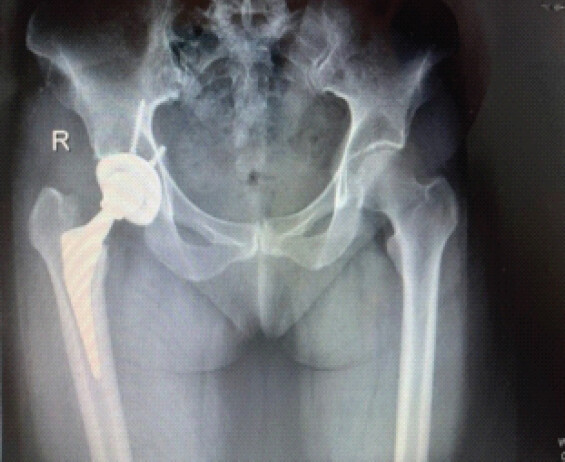
Repeat AP radiography of the hip demonstrated no obvious abnormalities. The acetabular component was positioned reasonably well, with slightly increased anteversion and inclination. The femoral stem appeared centered within the medullary canal, with no evidence of subsidence or fracture. A small gap between the prosthesis and the lateral cortex was noted, but proximal and medial fit appeared acceptable. The relatively short stem length was initially attributed to implant design (Fig. 1).
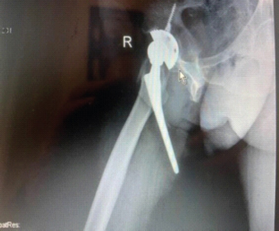
However, the lateral radiograph revealed a catastrophic finding: the femoral stem had perforated the posterior cortex and protruded posteromedially just inferior to the lesser trochanter. The stem tip was tenting the skin, producing a visible and palpable protuberance on the posteromedial thigh (Fig. 2).
The diagnosis and treatment options were explained to the patient, and revision surgery was strongly recommended. She elected to return to her primary surgeon and declined revision surgery at our institution.
Discussion
Proximal longitudinal fractures during uncemented hip arthroplasty are not uncommon and have been reported to occur in approximately 0.58% of primary hip arthroplasties; however, femoral cortical penetration is rare, with an incidence of only 0.07% [9]. Intraoperative femoral perforation during THA usually occurs during femoral canal preparation or insertion of the femoral component. Wade et al. identified two main risk factors for femoral perforation: Abnormal proximal femoral anatomy and difficulty with surgical exposure [7]. A BMI >40 has been defined as a criterion for difficult exposure.
In the present case, the patient had no history of previous hip surgery or proximal femoral deformity that could have led to erroneous stem guidance and subsequent femoral perforation. The patient’s BMI was 28, effectively excluding obesity-related difficulty with exposure. Posterior femoral perforation is not unexpected when an anterolateral approach is used. A broach starting point too close to the anterior cortex may redirect the instrument posteriorly. In addition, excessive lateralization near the greater trochanter in an attempt to avoid varus implantation may create an anterolateral-to-posteromedial force vector, predisposing the femur to posteromedial cortical perforation.
Osteoporosis and poor bone quality are associated with reduced cortical thickness and an increased risk of fracture. Patients with osteoporosis have been shown to have a 2.4-fold higher risk of fracture compared with those without osteoporosis [10]. The canal-to-bone ratio is a validated method for assessing proximal femoral bone quality on plain radiographs [11]. Based on this ratio, Dorr et al. classified proximal femoral morphology into three types, with Dorr type C femurs demonstrating a two- to five-fold increased risk of intraoperative periprosthetic fracture [12]. Although bone mineral density was not measured in the present patient, proximal femoral morphology was consistent with Dorr type A, making severe osteoporosis and a high fracture risk unlikely.
Most cases of femoral perforation complicating THA are detected either intraoperatively or in the early post-operative period [4,5,6,7]. Cortical perforation secondary to redirection of the femoral stem due to a previously implanted fibular strut graft has been reported [6]. In that case, hip instability with easy dislocation raised suspicion, and intraoperative posteromedial femoral stem perforation was confirmed using portable radiographs; revision surgery was performed after removal of both the stem and the fibular strut graft. In a retrospective study of 3,973 THAs, 16 patients (0.4%) sustained femoral perforation during broaching, all of which were diagnosed intraoperatively [4]. Wade et al. reported four cases of femoral perforation, all detected early due to severe pain during ambulation, and all were revised before plan hospital discharge [7]. In all reported cases, AP radiographs appeared normal, and the diagnosis was established exclusively on lateral radiographs.
In the present case, femoral perforation occurred despite the absence of recognized predisposing factors such as obesity, severe osteoporosis, or previous femoral deformity. Surgeons should therefore remain vigilant for this potential complication in all cases. Palpation of the cortical walls using an intramedullary suction tip or curette following broaching may facilitate early detection of cortical violation. When there is any intraoperative doubt, two-view radiographic evaluation should be obtained. In this case, the patient was able to ambulate with support for 15 days postoperatively and was evaluated by two orthopedic specialists; however, femoral perforation was overlooked due to reliance on the patient’s ability to ambulate and the normal appearance of AP radiographs. This case underscores the importance of routine two-view radiographic assessment in patients undergoing THA, particularly those presenting with persistent pain, regardless of the presence of classic risk factors.
Conclusion
Femoral cortical perforation during THA is a rare but potentially catastrophic complication that may remain unrecognized despite preserved ambulation and unremarkable AP radiographs. This case highlights that the absence of classical risk factors does not preclude the occurrence of femoral perforation. Surgeons should maintain a high index of suspicion in patients presenting with persistent or atypical post-operative thigh pain. Routine use of two-view radiographic evaluation, including lateral imaging, is essential both intraoperatively when doubt exists and postoperatively to ensure early diagnosis and prevent serious morbidity.
Clinical Message
Persistent or atypical thigh pain after THA should raise suspicion for femoral cortical perforation, even in patients who are able to ambulate and have normal AP radiographs. Routine use of two-view radiographic evaluation, including a lateral view, is essential for the early diagnosis and prevention of catastrophic complications.
Conflict of Interest:
Nil
Source of Support:
Nil
Consent
The authors confirm that informed consent was obtained from the patient for publication of this article
How to Cite this Article
Yıldırım Y, Gokkus K, Msangjı WJ. Late Diagnosis of Femoral Shaft Perforation During Total Hip Arthroplasty: Case Report. Journal of Orthopaedic Case Reports 2026 October;16(10): 219-222.
References
- Doyle J, Procter P, Moloney MA. Femoral shaft perforation at arthroplasty: To treat or not to treat. Arch Orthop Trauma Surg 1990;109:217-20. [Google Scholar] | [PubMed]
- Dammerer D, Putzer D, Glodny B, Petersen J, Arrich F, Krismer M. Occult intra-operative periprosthetic fractures of the acetabulum may affect implant survival. Int Orthop 2019;43:1583-90. [Google Scholar] | [PubMed]
- Pellici PM, Inglis AE, Salvati EA. Perforation of the femoral shaft during total hip replacement. J Bone Joint Surg Am 1980;62:234-40. [Google Scholar] | [PubMed]
- Kinney MC, Ho H, Hamilton WG. AAHKS best podium presentation research award: Femoral perforation during direct anterior approach total hip arthroplasty: Incidence, cohort characteristics, and management. J Arthroplasty 2022;37:S721-6. [Google Scholar] | [PubMed]
- Talmo CT, Bono JV. Preventing and managing intraoperative fractures and perforations in hip arthroplasty. Orthopedics 2005;28:s1085-8. [Google Scholar] | [PubMed]
- Huda N, Julfiqar, Pant A, Aslam M. An unexpected complication of residual fibular strut graft while performing a press fit hemiarthroplasty for a secondary nonunion fracture neck of the femur. J. Orthop Case Rep 2015;5:45-7. [Google Scholar] | [PubMed]
- Wade FA, Parvizi J, Sharkey PF, Purtill JJ, Hozack WJ. Femoral perforation complicating contemporary uncemented hip arthroplasty. J Arthroplasty 2006;21:452-5. [Google Scholar] | [PubMed]
- Siddiqi A, Springer BD, Chen AF, Piuzzi NS. Diagnosis and management of intraoperative fractures in primary total hip arthroplasty. J Am Acad Orthop Surg 2021;29:e497-512. [Google Scholar] | [PubMed]
- Parvizi J, Rapuri VR, Purtill JJ, Sharkey PF, Rothman RH, Hozack WJ. Treatment protocol for proximal femoral periprosthetic fractures. J Bone Joint Surg 2004;86-A Suppl 28-16. [Google Scholar] | [PubMed]
- Sun G, Yin Y, Ye Y, Li Q. Risk factors for femoral fracture in lateral decubitus direct anterior approach total hip arthroplasty using conventional stems: A retrospective analysis. J Orthop Surg Res 2021;16:98. [Google Scholar] | [PubMed]
- Yeung Y, Chiu KY, Yau WP, Tang WM, Cheung WY, Ng TP. Assessment of the proximal femoral morphology using plain radiograph-can it predict the bone quality?. J Arthroplasty 2006;21:508-13. [Google Scholar] | [PubMed]
- Dorr LD, Faugere MC, Mackel AM, Gruen TA, Bognar B, Malluche HH. Structural and cellular assessment of bone quality of proximal femur. Bone 1993;14:231-42. [Google Scholar] | [PubMed]
© 2026 Journal of Orthopaedic Case Reports - Published by Indian Orthopaedic Research Group
About the Authors
-

Yakup Yıldırım
-

Kemal Gokkus
-

Waziri Juma Msangı
Related Articles in Journal of Orthopaedic Case Reports
 October 1, 2026 Total Hip Arthroplasty for Neglected Pathological Femoral Neck Fracture in a Young Patient with Autoimmune Polyglandular Syndrome
 October 1, 2026 A Study of Spinopelvic Relationship and its Impact on Functional Outcome of Total Hip Arthroplasty
 October 1, 2026 Primary Total Hip Arthroplasty for a Displaced Femoral Neck Fracture in an Above-the-Knee Amputee: A Case Report
 October 1, 2026 Total Hip Arthroplasty for Advanced Femoral Head Osteonecrosis in a 24-Year-Old Competitive Wrestler: A Case Report