


Introduction
Meniscal abnormalities are rare but can cause various symptoms, such as pain, swelling, and locking [1]. Among these, the discoid meniscus is the most common, with an incidence of 0.4–17%, whereas the double-layered lateral meniscus is extremely rare, accounting for <1% of all meniscal anomalies [2]. Although there is no strict definition of double-layered meniscus, it is generally considered as the presence of an accessory meniscus that exists separately from the normal meniscus, originates near the anterior or posterior horn attachment of the normal meniscus, and attaches to the lateral or medial femoral condyle. Although arthroscopic meniscectomy has been reported as an effective treatment for the double-layered lateral meniscus, its characteristic magnetic resonance imaging (MRI) findings remain unclear, and arthroscopic observation provides the definitive diagnosis [3].
In this report, we present a 47-year-old man with left knee pain. MRI initially suggested a locked lateral meniscus, but arthroscopy revealed double layers of the lateral meniscus. The patient was successfully treated with arthroscopic meniscectomy.
Case Report
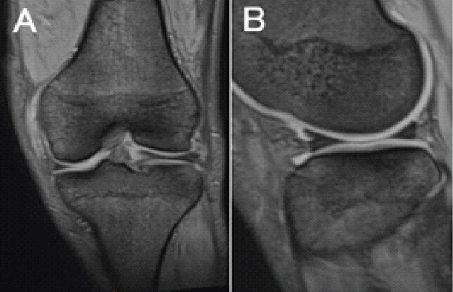
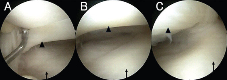
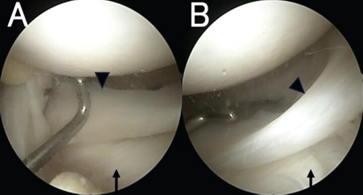
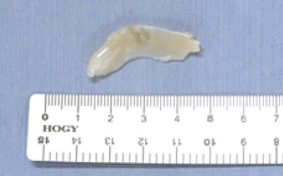
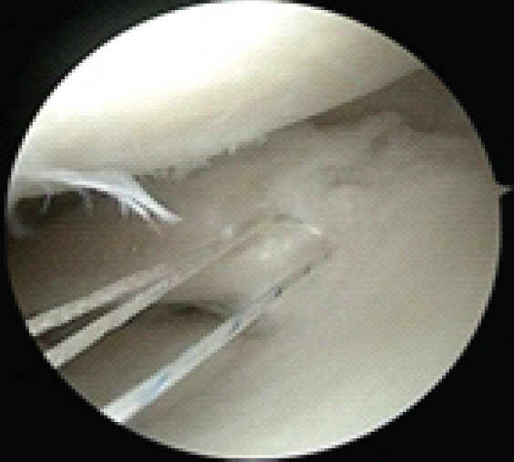
A 47-year-old male presented to our department with left knee pain during martial arts training. Physical examination revealed no swelling or patellar ballottement, with a range of motion from 0° extension to 140° flexion. Both the McMurray test and Lachman test were negative, as was the sagging sign. Plain radiographs revealed no abnormal findings, including bone injury or osteoarthritic changes. Coronal MRI suggested lateral meniscal locking, with a duplicated meniscal structure overlapping the native lateral meniscus attached to the tibia. In the sagittal plane, thickening and signal changes on T2-weighted images were observed in the anterior horn of the lateral meniscus. However, the inner margin of the anterior horn appeared sharp on the sagittal view, making it difficult to definitively diagnose a meniscal tear (Fig. 1). An arthroscopic examination was performed for diagnostic and therapeutic purposes, revealing a double-layered lateral meniscus with the superior layer flipped anteriorly (Fig. 2). On the double-layered meniscus, the unstable superior layer extended from the anterior horn to the midbody (Fig 3). This superior layer was resected entirely (Fig. 4). A horizontal tear was observed in the inferior meniscus after resection of the superior layer; this was repaired using an all-inside device (JuggerStitch Meniscal Repair Device; Zimmer-Biomet, Warsaw, IN) (Fig. 5). Post-repair, stability of the meniscus was confirmed. Partial cartilage damage was found on the lateral tibial plateau. The procedure was completed without major complications. Post-operatively, range-of-motion exercises and weight-bearing ambulation were initiated according to pain tolerance.
Discussion
In the present case, symptom improvement was confirmed after the resection of the accessory meniscal layer. Furthermore, we found that MRI may serve as an adjunctive tool for pre-operative assessment of the double-layered meniscus.
This case showed that resection of the accessory meniscus resulted in symptomatic improvement. The double-layered lateral meniscus is a rare morphological abnormality of the meniscus. Ryu et al. conducted an anatomical study of 437 patients and reported that 0.5% of meniscal abnormalities were double-layered lateral meniscus [4]. A double-layered meniscus is known to cause pain, swelling, and mechanical locking of the knee. Although a partial tear of the lateral meniscus was identified in the present case, clinical findings strongly suggestive of a symptomatic meniscal tear – such as joint swelling or a definitively positive McMurray test – were absent. Arthroscopic observation is the definitive diagnosis and most previously reported cases of the double-layered lateral meniscus underwent meniscectomy; our case similarly demonstrated symptom resolution following resection [1,5,6,7,8]. While partial horizontal tears in the inferior native lateral meniscus were repaired using the all-inside device, it remains unclear whether these tears were directly related to the double-layered meniscus.
This case suggested that an accessory meniscal layer can be suspected on MRI when the inner margin of the meniscus appears sharp, and when anterior or posterior horns are observed without tibial attachment. As characteristic MRI findings for the double-layered lateral meniscus have not yet been established, definitive diagnosis typically requires arthroscopic confirmation of a double-layered meniscal structure. Ota et al. reported that sagittal MRI images revealed signal changes and thickening within the meniscus, while coronal images suggested intercondylar locking typically associated with discoid meniscus tears [3]. Shimozaki et al. described posterior horn thickening as a feature of inverted-type discoid meniscus tears; however, whereas the inner edge of the meniscus appears blunted in such cases, the double-layered lateral meniscus observed in the present case showed a sharp inner edge [9]. Although the coronal appearance may mimic intercondylar locking of a discoid meniscus, this finding in the double-layered lateral meniscus is attributed to the presence of anterior and posterior horns that do not attach to the tibia, in addition to a normally attached meniscus.
Conclusion
Resection of the accessory meniscal layer can improve symptoms in patients with a double-layered lateral meniscus. It could be suspected on MRI by the presence of sharp inner meniscal margins and non-attached anterior/posterior horns. When such MRI findings are present, the double-layered lateral meniscus should be considered in the differential diagnosis, and meniscectomy may provide symptomatic relief.
Clinical Message
Double-layered lateral meniscus can mimic meniscal locking on MRI. Recognition of sharp inner margins and non-attached horns on MRI may help preoperative suspicion and arthroscopic resection yields good results.
Conflict of Interest:
Nil
Source of Support:
Nil
Consent:
The authors confirm that informed consent was obtained from the patient for publication of this article
How to Cite this Article
Katsuo T, Takata Y, Takemoto N, Nishimura M, Demura S, Nakase J. Double-Layered Lateral Meniscus Mimicking Locking of Lateral Meniscus: A Case Report. J Orthop Case Rep. 2026 November;16(11): 126-129. doi: 10.13107/jocr.2026.v16.i11.8412.
References
- Oklaz EB, Kaya I, Ciceklidag M, Bircan R, Tosun MF, Kanatli U.. Discoid double-layered lateral meniscus: A case report. J Orthop Case Rep 2024;14:152-5. [Google Scholar] | [PubMed]
- Okahashi K, Sugimoto K, Iwai M, Oshima M, Fujisawa Y, Takakura Y.. Double-layered lateral meniscus. J Orthop Sci 2005;10:661-4. [Google Scholar] | [PubMed]
- Ota M, Takagi H, Kato S, Kawashima F, Kanzaki K.. Surgical treatment of double-layered lateral meniscus. Case Rep Orthop 2021;2021:9978889. [Google Scholar] | [PubMed]
- Ryu K, Iriuchishima T, Oshida M, Saito A, Kato Y, Tokuhashi Y. Evaluation of the morphological variations of the meniscus: A cadaver study. Knee Surg Sports Traumatol Arthrosc 2015;23:15-9. [Google Scholar] | [PubMed]
- Ahn TY, Kim HT, Park YG.. Double-layered lateral meniscus including a ring-shaped accessory meniscus: A case report. JBJS Case Connect 2018;8:e104. [Google Scholar] | [PubMed]
- Looney AM, Ferris WJ, West RV.. Rare variant double-layered lateral meniscus detected during arthroscopy in a young gymnast after a hyperextension knee injury: A case report. JBJS Case Connect 2020;10:e1900648. [Google Scholar] | [PubMed]
- Wang Q, Liu XM, Liu SB, Bai Y.. Double-layered lateral meniscus. Knee Surg Sports Traumatol Arthrosc 2011;19:2050-1. [Google Scholar] | [PubMed]
- Araki S, Kubo M, Kumagai K, Imai S.. Double-layered lateral meniscus in an 8-year-old child: Report of a rare case. Case Rep Orthop 2016;2016:5263248. [Google Scholar] | [PubMed]
- Shimozaki K, Nakase J, Takata Y, Asai K, Toyooka K, Kitaoka K. The characteristic findings of an inverted-type discoid lateral meniscus tear: A hidden tear pattern. BMC Musculoskelet Disord 2019;20:223. [Google Scholar] | [PubMed]
© 2026 Journal of Orthopaedic Case Reports - Published by Indian Orthopaedic Research Group
About the Authors
-

Takashi Katsuo
-

Yasushi Takata
-

Naoki Takemoto
-

Manase Nishimura
-

Satoru Demura
-

Junsuke Nakase
Related Articles in Journal of Orthopaedic Case Reports
 November 1, 2026 Incidentally Detected Osseous Hemangioma of the Femoral Head: Conservative Management of a Rare Case with Review of the Literature
 October 1, 2026 Magnetic Resonance Imaging-Arthroscopy Correlation in Traumatic Knee Injuries: A Retrospective Diagnostic Accuracy Study
 October 1, 2026 Incidence, Arthroscopic Detection, and Clinical Outcomes of Ramp Lesions in Patients Undergoing Anterior Cruciate Ligament Reconstruction: A Prospective Observational Study
 October 1, 2026 Acute Cauda Equina Syndrome in Rapid Succession: A Case Series of Eight Consecutive Patients Undergoing Emergency Surgical Decompression Over One Week