

In patients with hip and spinal ankylosis, total hip arthroplasty through the direct anterior approach in the supine position, aided by fluoroscopy and dual-mobility components, minimizes instability and facilitates early rehabilitation despite severe pre-operative deformity.
Dr. Chandra Kumar Khande, Department of Orthopaedics, Institute of Medical Sciences, Banaras Hindu University, Varanasi, Uttar Pradesh, India. E-mail: chandrakumar.khandey@gmail.com
Abstract
Introduction: Bony ankylosis of the hip results in profound functional disability and poses significant challenges during total hip arthroplasty (THA), especially when associated with spinal ankylosis. Altered anatomy, severe soft-tissue contractures, fixed deformities, and disturbed spinopelvic mechanics complicate patient positioning, anesthetic management, and component placement and increase the risk of post-operative instability. Although THA has demonstrated favorable functional outcomes in ankylosed hips, limited literature highlights the role of the direct anterior approach (DAA) in patients with concomitant spinal fusion. We report a rare case of bilateral hip and spinal ankylosis managed successfully using DAA THA.
Case Report: A 40-year-old female, completely bedridden for 10 years, presented with the inability to sit, stand, or perform activities of daily living due to long-standing stiffness of multiple joints. Clinical examination revealed complete ankylosis of both hips and the spine, with a 30° fixed flexion deformity of the left hip. Radiographs confirmed bony ankylosis of both hips and the entire spine. The Harris Hip Score was 0. Staged bilateral THA was performed using the DAA in the supine position under general anesthesia. Intraoperative fluoroscopy facilitated controlled acetabular reaming and accurate component positioning. Dual mobility cups were used to mitigate instability risk related to absent spinopelvic motion. Early post-operative mobilization was initiated. At the 1-year follow-up, the patient achieved independent sitting and assisted ambulation, with the Harris Hip Score improving to 80 and no episodes of dislocation or major complications.
Conclusion: DAA THA is a feasible and effective option in patients with ankylosed hips and spines. Supine positioning and fluoroscopic guidance enhance surgical accuracy and stability, enabling significant functional recovery.
Keywords: Ankylosing spondylitis, fused hip, total hip arthroplasty, direct anterior approach, dual mobility.

Bony ankylosis of the hip may arise secondary to chronic inflammatory disorders such as ankylosing spondylitis or psoriatic arthritis, or it may occur as a long-term consequence of infection or trauma. Although the hip in these cases is often painless, the resultant loss of motion severely restricts daily activities. Inflammatory etiologies may further compound disability through concomitant spinal stiffness and polyarticular involvement. Total hip arthroplasty (THA) is widely accepted as the treatment of choice for restoring mobility and improving quality of life in this patient population. Previous studies have consistently demonstrated marked functional gains and high patient satisfaction following conversion of an ankylosed hip to THA [1]. However, despite these favorable outcomes, reports seldom emphasize the technical and perioperative difficulties encountered in such cases. Challenges arise as early as patient positioning on the operating table, particularly in the presence of fixed deformities of the hip, spine, or knee, and extend to intraoperative exposure, implant placement, and early post-operative mobilization [2]. These cases also pose significant challenges for anesthesiologists, particularly due to ankylosed spines, spinal stiffness, and difficulties with positioning and airway management. Furthermore, these patients are predisposed to dislocation, largely due to the risk of component malpositioning in the setting of distorted anatomy [3]. Historically, THA in ankylosed hips has been performed through posterior or lateral approaches with the patient positioned laterally. More recently, the direct anterior approach (DAA) has gained popularity for primary and complex THA, primarily due to its muscle-sparing technique, comfortable patient position, and the ability to utilize intraoperative imaging for accurate component placement [4,5]. Given these potential advantages, we present a case report of a completely bedridden patient with ankylosis of both hip and spine operated via DAA.
A 40-year-old female, a completely bedridden lady, presented with complaints of multiple joint stiffness and the inability to sit or stand for 10 years (Fig. 1).
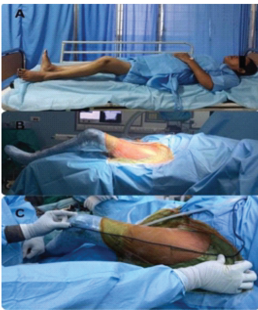
Figure 1: Pre-operative clinical images of the patient, (a) bedridden patient with fixed flexion deformity at the hip joint, (b) supine patient positioning for direct anterior approach, (c) marking of incision line.
She was unable to perform her basic daily routine activities, such as maintenance of personal hygiene, sitting on a chair, and washing clothes. She was also not sexually active for the last 10 years. The clinical examination revealed no movement in both hip joints and the spine, with a fixed flexion deformity of 30° in the left hip. On X-ray, bony ankylosis of both hips, along with the whole spine, was present (Fig. 2).
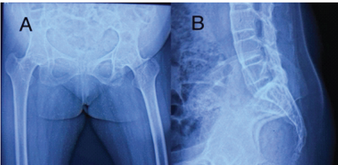
Figure 2: Pre-operative radiographs. (a) Pre-operative radiograph of pelvis with both hip (anteroposterior view), (b) pre-operative radiograph of lumbosacral spine (lateral view).
The sacral slope and lumbar lordosis of the patient were 36° and 38°, respectively. As the patient was completely bedridden, assessment of the change in spinopelvic parameters with patient position could not be done, and determining the type of hip–pelvis relationship was difficult. The Harris Hip Score was zero. Staged THA of the bilateral hip via DAA was planned.
Surgical technique:
The patient was positioned supine on a standard operating table under general anesthesia. A longitudinal skin incision of approximately 8–10 cm was made along a line drawn from a point 2.5 cm distal and lateral to the anterior superior iliac spine toward the fibular head. After subcutaneous dissection, a longitudinal incision was made over the anterior fascia of the tensor fascia lata (TFL). After fascial incision, two flaps were developed. The medial flap was retracted with Allis forceps, and blunt dissection beneath the TFL exposed the posterior fascia. Small ascending branches of the lateral circumflex femoral artery encountered during this step were coagulated.
Acetabular preparation (Fig. 3):
- Retraction of the TFL laterally provided access to the anterior capsule of the hip
- Curved retractors were placed to expose the capsule and anterior column
- A T-shaped capsulotomy was created to visualize the femoral neck and ankylosed joint
- Femoral neck osteotomy was performed to excise a napkin-ring-like bone fragment (Fig. 2)
- The proximal femur was mobilized posteriorly using a Mueller retractor
- Acetabular reaming was started with the smallest reamer under fluoroscopic guidance and progressed till the appropriate size
- Cementless, press-fit acetabular cups were seated and fixed with appropriately sized screws.
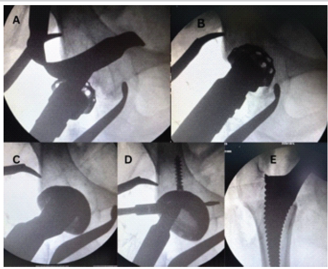
Figure 3: Intraoperative C-arm images of a patient. (a and b) Sequential reaming, (c) acetabular component placement, (d) acetabular component fixation with screws, (e) Trial femoral stem placement and trial reduction.
Femoral preparation (Fig. 3):
- A capsulo-labral release was carried out at the 1 o’clock position for the left hip and at 11 o’clock for the right hip
- The inferior capsule was released along the femoral neck to the lesser trochanter, with the limb externally rotated
- Superior capsule release at the trochanteric fossa was performed with femoral elevation and anterior translation
- The operating table was adjusted to extend and adduct the hip, which facilitated broaching
- Sequential broaching was followed by trial reductions to confirm stability and limb length. Final femoral implants were inserted under fluoroscopic guidance.
Closure and post-operative management:
The anterior TFL fascia was repaired with Vicryl sutures, followed by layered closure of subcutaneous tissue and skin. Post-operative mobilization and range of motion exercises were initiated on the 2nd post-operative day, beginning with bedside sitting and progressing to ambulation as tolerated. Indomethacin was prescribed for 6 weeks to reduce the risk of heterotopic ossification. The patient was followed up for 1 year, and her Harris Hip Score improved to 80 (Fig. 4).
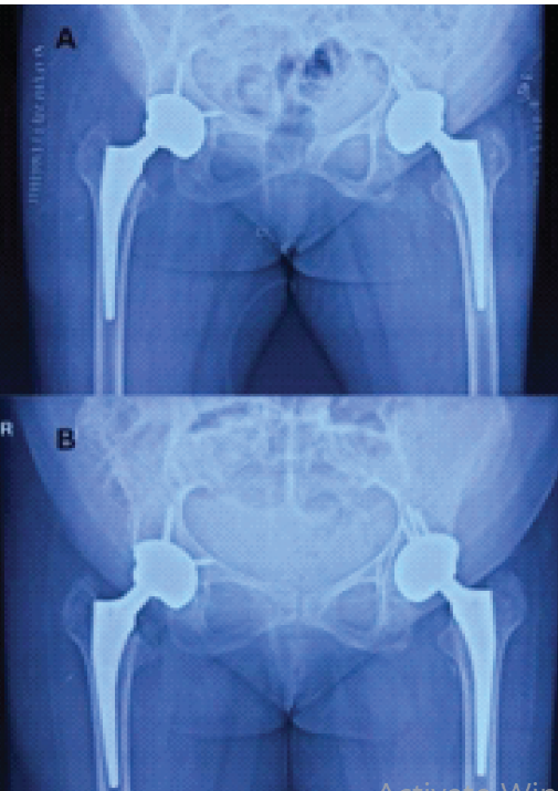
Figure 4: Post-operative radiographs of the pelvis with both hip (anteroposterior view). (a) Immediate post-operative radiograph, (b) radiograph at 1-year follow-up.
THA can produce dramatic improvements in pain, function, and overall quality of life for patients with bony ankylosis of the hip. However, these procedures are technically demanding. Structural deformities of the pelvis and proximal femur, soft-tissue contractures, and disuse osteopenia increase the complexity of exposure and component placement and raise the risk of intraoperative and post-operative complications [3]. The creation of a new acetabular bed in a previously fused joint and the careful handling of fragile bone make surgical planning and intraoperative technique particularly crucial. Historically, a variety of surgical approaches have been utilized for THA in ankylosed hips, including lateral approaches with trochanteric osteotomy, posterior approaches, and combined anterior–posterior strategies. Each approach has recognized advantages, but none is without limitations; common challenges across techniques include limited visualization, risk of neurovascular injury, higher blood loss, and potential for iatrogenic fracture. Moreover, malpositioning of components, especially the acetabular cup, can result from distorted landmarks and inadequate intraoperative imaging, contributing to instability and suboptimal biomechanics. Anticipated problems in this case were:
- Risk of fracture of the spine while shifting the patient to the operating table [6]
- Difficult neuraxial anesthesia and airway management difficulties due to a fused spine
- Difficult patient positioning, especially the lateral position, due to the presence of fixed flexion and abduction deformities of the hip
- Limited exposure, component malpositioning, and difficult intraoperative imaging
- Increased risk of post-operative dislocation.
We adopted the DAA with the patient in the supine position to address several of these problems. A Taylor’s brace was applied before shifting the patient to the operating table to reduce the chances of vertebral fracture. Video-assisted laryngoscopy backup was available to manage a difficult airway. DAA is a true inter-nervous and inter-muscular approach that facilitates enhanced recovery after surgery. Performing the procedure supine permits reliable use of intraoperative fluoroscopy, which aids controlled acetabular reaming and precise orientation of the cup [7]. Real-time imaging thereby helps avoid inadvertent over-reaming, malalignment, and unrecognized leg-length discrepancy. Intraoperative imaging, leading to precise component positioning and muscle preservation, leading to early recovery, significantly reduces the chances of postoperative dislocation [8,9]. Normally, when a person changes his posture from standing to sitting, movement occurs at the hips, spinopelvic junction, and lumbar spine. Moreover, in our patient, as the spinopelvic junction and spine were completely fused, more motion would be required at the hip during a change of posture, which in turn would lead to the risk of anterior impingement and posterior dislocation [10,11]. To counteract this challenge, we increased acetabular anteversion and used a dual mobility cup, which can provide more range of motion at the hip [12]. In this case, the patient experienced rapid pain relief and early mobilization, resulting in shorter hospital stays and no early dislocations. These outcomes are consistent with the theoretical benefits of a muscle-preserving approach combined with image-assisted implantation of a dual mobility cup. Sciatic nerve palsy is a known complication reported with posterior approaches, either from direct trauma or from a stretch during intraoperative limb lengthening. The supine DAA allows intraoperative clinical and radiographic assessment of limb length and offset before final component fixation, reducing the likelihood of unrecognized vertical offset changes that could stretch the sciatic nerve [13]. After 1 year of follow-up, the patient had a satisfactory improvement in functional outcome with a significant improvement in her Harris Hip Score (0–80) (Fig. 5). As the patient was also having ankylosis of the spine and knee pain, these factors led to a decrease in the Harris Hip Score on final follow-up. We present a rare case with grossly fused hip joints as well as the spine, which required very precise pre- and intraoperative planning. DAA in an ankylosed hip needs surgical expertise and a longer learning curve.
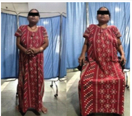
Figure 5: Post-operative clinical images of the patient.
The DAA for THA in patients with bony ankylosis, although technically demanding, is a feasible and effective option. Supine positioning and intraoperative imaging enhance surgical feasibility. Faster post-operative functional recovery, shorter hospitalization, and comparatively fewer chances of dislocation suggest potential advantages of DAA over other approaches.
DAA THA with intraoperative fluoroscopic guidance and dual mobility implants is a safe and effective option for managing ankylosed hips with spinal fusion, allowing accurate component placement and early functional recovery.
References
- 1. Bukowski BR, Clark NJ, Taunton MJ, Freedman BA, Berry DJ, Abdel MP. Primary total hip arthroplasty in patients with ankylosing spondylitis. J Arthroplasty 2021;36:S282-9. [Google Scholar] [PubMed]
- 2. Blizzard DJ, Penrose CT, Sheets CZ, Seyler TM, Bolognesi MP, Brown CR. Ankylosing spondylitis increases perioperative and postoperative complications after total hip arthroplasty. J Arthroplasty 2017;32:2474-9. [Google Scholar] [PubMed]
- 3. Mekkawy KL, Rodriguez HC, Mercadal G, Gosthe RG, Khanuja HS, Corces A, et al. Ankylosing spondylitis in patients undergoing total hip arthroplasty increases the risk of medical and implant-related complications: A case control analysis. Hip Int 2024;34:602-7. [Google Scholar] [PubMed]
- 4. Flevas DA, Tsantes AG, Mavrogenis AF. Direct anterior approach total hip arthroplasty revisited. JBJS Rev 2020;8:e0144. [Google Scholar] [PubMed]
- 5. Ang JJ, Onggo JR, Stokes CM, Ambikaipalan A. Comparing direct anterior approach versus posterior approach or lateral approach in total hip arthroplasty: A systematic review and meta-analysis. Eur J Orthop Surg Traumatol 2023;33:2773-92. [Google Scholar] [PubMed]
- 6. Mahesh BH, Jayaswal A, Bhan S. Fracture dislocation of the spine after total hip arthroplasty in a patient with ankylosing spondylitis with early pseudoarthrosis. Spine J 2008;8:529-33. [Google Scholar] [PubMed]
- 7. Chen P, Liu W, Wu C, Ruan P, Zeng J, Ji W. Fluoroscopy-guided direct anterior approach total hip arthroplasty provides more accurate component positions in the supine position than in the lateral position. BMC Musculoskelet Disord 2023;24:884. [Google Scholar] [PubMed]
- 8. Christensen TH, Egol A, Pope C, Shatkin M, Schwarzkopf R, Davidovitch RI, et al. How does surgical approach affect characteristics of dislocation after primary total hip arthroplasty? J Arthroplasty 2023;38 Suppl 2:S300-5. [Google Scholar] [PubMed]
- 9. Haynes JA, Hopper RH Jr., Ho H, McDonald JF, Parks NL, Hamilton WG. Direct anterior approach for primary total hip arthroplasty lowers the risk of dislocation compared to the posterior approach: A single institution experience. J Arthroplasty 2022;37:495-500. [Google Scholar] [PubMed]
- 10. Oommen AT, Hariharan TD, Chandy VJ, Poonnoose PM, Arun Shankar A, Kuruvilla RS, et al. Total hip arthroplasty in fused hips with spine stiffness in ankylosing spondylitis. World J Orthop 2021;12:970-82. [Google Scholar] [PubMed]
- 11. Haffer H, Adl Amini D, Perka C, Pumberger M. The impact of spinopelvic mobility on arthroplasty: Implications for hip and spine surgeons. J Clin Med 2020;9:2569. [Google Scholar] [PubMed]
- 12. Hussein Y, Iljazi A, Sørensen MS, Overgaard S, Petersen MM. The risk of dislocation in dual-mobility versus 36 mm heads in primary total hip arthroplasty for osteoarthritis. Bone Jt J 2025;107-B:50-7 [Google Scholar] [PubMed]
- 13. Slaven SE, Ho H, Sershon RA, Fricka KB, Hamilton WG. Motor nerve palsy after direct anterior versus posterior total hip arthroplasty: Incidence, risk factors, and recovery. J Arthroplasty 2023;38:S242-6. [Google Scholar] [PubMed]






Related Articles in Journal of Orthopaedic Case Reports
 July 1, 2026 Managing Complications of Total Hip Arthroplasty for Avascular Necrosis Hip Performed Via Anterior Approach
 May 1, 2026 Direct Anterior THA with Leg Positioning Traction System for Avascular Necrosis: Technique and Early Outcomes
 March 1, 2026 Day-Care Direct Anterior Total Hip Arthroplasty in Urban India: Patient Education, Apprehensions, and Outcomes
 March 1, 2026 Clinical and Safety Outcomes after Direct Anterior Total Hip Arthroplasty Compared with Conventional Approaches: A Systematic Review and Meta-Analysis