


Introduction
Heterotopic ossification (HO) is a benign ossification of soft tissues, categorized into three types: genetic, traumatic, and neurogenic HO [1,2,3].
HO is a frequent complication following trauma, occurring in 10–20% of patients with traumatic brain injury (TBI) or spinal cord injury (SCI), and up to 30–50% in patients with fractures [4,5]. Prolonged mechanical ventilation has additionally been identified as a risk factor for severe HO [6]. Underlying mechanisms include a pro-inflammatory environment with overexpression of osteogenic mediators like bone morphogenetic protein (BMP)-2 and BMP-4, which can cause pathological endochondral ossification in HO-susceptible locations [5]. The most commonly affected joints include the hip, elbow and knee, although HO can develop almost anywhere in the body [7,8]. HO presenting specifically within muscle tissue is classified as myositis ossificans [9,10,11].
HO typically presents 3 to 12 weeks after an inciting event with tissue injury. Clinical signs include increased joint stiffness, limited range of motion, warmth, swelling, and erythema [2,12]. As these clinical signs are often non-specific, diagnosis is commonly made with plain radiographs and computed tomography, showing ectopic bone fragments with peripheral ossification zones.
Other imaging techniques include ultrasound and bone scintigraphy [5,8]. Early ultrasound findings include a hypoechoic core with an outer hyperechoic rim, while bone scintigraphy typically shows increased uptake in the injured area [2,13]. Due to the initial lack of bone formation, plain radiographs may not show any abnormalities in the first 2 weeks after injury, with ultrasound and bone scintigraphy having a higher sensitivity in the early stages [2,8]. Despite this, early diagnosis remains challenging due to the initial non-specific symptoms, and the lack of reliable tools or methods to predict which patients are likely to develop HO [14].
Evidence for the non-surgical treatment of HO is limited. Non-surgical care usually consists of physical therapy management and non-steroidal anti-inflammatory drugs (NSAIDs). This evidence is extrapolated from the effectiveness of NSAIDs in the prophylactic treatment after hip surgery [2]. Gentle mobilization of the joint within a pain-free range is recommended; forcible manipulation is contraindicated. It remains uncertain if physical therapy has a significant role in the development or reduction of HO [5,12].
Treatment with bisphosphonates is controversial, but they have been shown to be effective in some causes of HO, i.e., burn injuries and SCI [3,7,15].
Surgery should be considered in patients with functional deficits that failed conservative management. The timing for surgical excision is under discussion and is guided by the etiology and bone maturation. A waiting time of 6 months for maturation seems to be adequate in the post-traumatic setting without TBI or SCI. In SCI, HO is considered mature after 12 months and 18 months for TBI [2,4,8,12]. Recurrence rates remain high after surgery, making secondary prevention with NSAIDs and/or radiotherapy essential to reduce the risk of recurrence.
Case Report
We present the case of a male patient in his mid-twenties who was involved in a motor vehicle accident resulting in numerous fractures (left humerus, radius, ulna, ribs II-VII, femur condyle, patella; right mandible, ribs I-II-III, femur) and severe blunt abdominal trauma. Orthopedic surgical management encompassed osteosynthesis of the mandible, radius, ulna, intramedullary nail of the right femur, left humerus, and screw fixation of the left femoral condyle. The abdominal trauma required multiple laparotomies. The patient required 7 weeks of invasive mechanical ventilation in the intensive care unit.
Passive mobilization was started postoperatively with initial clinical improvement while the patient was still intubated. However, 4 weeks postoperatively, the patient presented with an increase in pain during mobilization and severe mobility regression in both knees and the right hip, alongside mild mobility loss in the left shoulder and elbow compared to 2 weeks beforehand.
Investigations
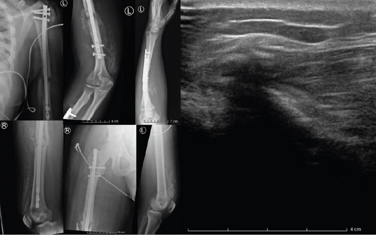
Plain radiographs and ultrasound revealed extensive HO in the left shoulder, forearm, and distal quadriceps muscles bilaterally (Fig. 1). In addition, the radiographs excluded displacement of the fractures or the orthopedic material.
Treatment
NSAIDs were initiated (naproxen 500 mg twice daily for 3 weeks) and further rehabilitation with progressive slow mobilization of the limbs.
After recovering sufficiently from his abdominal injuries, the patient commenced intensive inpatient rehabilitation consisting of five sessions per week of physical therapy and occupational therapy, each of at least 1 h/day, aimed at improving function, mobility and strength. This was coordinated by a multidisciplinary team including occupational therapists, physiotherapists, and physical medicine and rehabilitation physicians.
The option of surgical excision of the HO in combination with radiotherapy was offered at 8 months post-injury to increase mobility in the knees. Due to the extensiveness of the HO in both quadriceps muscles, surgery would result in significant muscle loss. The patient declined, reporting adequate functional recovery in activities of daily living despite the persistent mobility restrictions.
Outcome
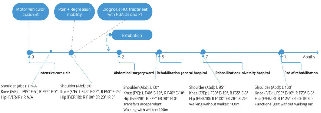
In total, the patient received 6 months of multidisciplinary rehabilitation which focused on mobility, strength, proprioception, functional training, and gait rehabilitation. An overview of the timeline is shown in Fig. 2.
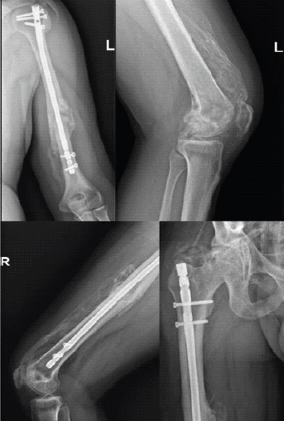
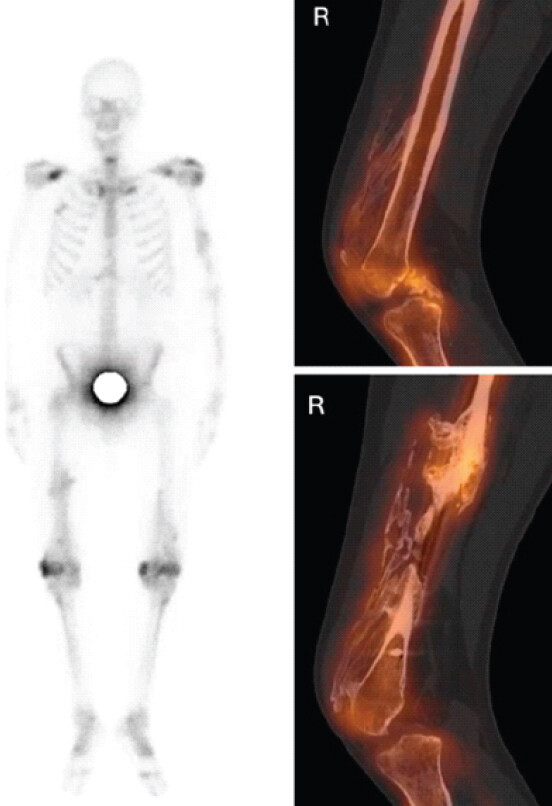
At 8 months follow-up, imaging with radiographs, ultrasound, and single-photon emission computed tomography scan revealed minimal bone turnover in the ossifications of the quadriceps with only mild tracer uptake distal anteriorly (Figs. 3 and 4). These signs of maturation supported the option to consider surgery.
After rehabilitation, the patient was able to regain near-normal walking speed without assistive devices; however, significant range of motion deficits persisted in both knees, with flexion limited to 55° (left) and 70° (right), and extension deficits of −10° (left) and −5°(right).
Discussion
HO is a known complication in polytrauma patients, though the incidence of multi-joint involvement without TBI or SCI is not well established. Older literature suggests that multi-joint involvement is seen in approximately 6% of head-injured patients [16]. HO with simultaneous involvement of the shoulder, forearm, hip, and both quadriceps, as seen in this case, is rare. To our knowledge, only one other case report of extensive multi-joint HO without head trauma has been reported, in a patient subjected to prolonged ventilation due to COVID-19 infection [17].
The pathophysiology of HO is still not fully understood. In traumatic HO, it is hypothesized that trauma causes inflammation, vascularization, and tissue ischemia at the site of injury. Through complex signaling pathways (including BMP-2, BMP-4), this leads to endochondral ossification, a process where cartilage is replaced by bone [5,7,8,18]. This is likely amplified by systemic factors. Wollner et al. [19] compared the epidemiology of HO in femur fractures treated with an intramedullary nail among polytrauma patients versus isolated femur fractures. HO was apparent in 75% of the polytrauma patients compared to 20% in the isolated fracture group. They hypothesized that the difference could be due to the hyperinflammatory state of the polytrauma patient and long immobilization. In addition, in polytrauma, there is frequent association with TBI and SCI, which are hypothesized to trigger osteogenic mediators (like Substance P, interleukin-6, BMPs, and fibroblast growth factors) [20]. Trauma type and energy also influence the risk, as the prevalence of HO in high-energy war trauma is far greater than in civilian injuries [21]. In our patient, prolonged mechanical ventilation likely further amplified the risk and extent of HO, consistent with previously reported cases [6,17].
There is limited evidence for the conservative management of HO. In this case, NSAIDs were administered as they have demonstrated significant efficacy in the prophylaxis of HO, as they inhibit Cyclooxygenase-2 which is required for endochondral ossification [4,5,22]. No direct therapeutic evidence for NSAIDs in established HO exists. Their use here aimed to limit further ossification. No consensus exists on optimal type, dosage or duration, and both selective and non-selective NSAIDs appear comparably effective for prophylaxis [8]. In patients with no contraindications to NSAIDs, the complication rates are relatively low, although an increased risk of non-union in acetabular fractures has been reported [23].
Given the high morbidity and rehabilitation burden associated with HO and lack of strong evidence for conservative management once HO has developed, early recognition and prophylaxis are crucial. In our case, HO necessitated multiple months of rehabilitation to improve function, but significant mobility restrictions persisted. It is our view that NSAID prophylaxis for a minimum of 2 weeks can be considered in polytrauma patients if there are no contraindications [22]. If NSAIDs are contraindicated, prophylaxis with radiotherapy, a single dose of 700 cGy from 24 h pre- to 72 h postoperatively, can be considered at high-risk locations, though this evidence is extrapolated from the arthroplasty setting [8,19,24].
Ultrasound can aid screening and early detection, as it is a cost-effective tool with higher sensitivity than plain radiographs in the early stage of HO [21]. Early recognition might be achieved using ultrasound screening at 1–2 weeks at high-risk HO locations (e.g., hips and knees) [25].
Conclusion
This case documents a rare presentation of extensive post-traumatic HO with simultaneous involvement of the left shoulder, forearm, right hip, and bilateral quadriceps, in a polytrauma patient without TBI or SCI. Prolonged mechanical ventilation likely contributed to the severity and distribution of ossification. Although 6 months of multidisciplinary rehabilitation enabled the patient to achieve near-normal ambulation, clinically significant knee range-of-motion deficits persisted at follow-up. This case underscores that early ultrasound screening at high-risk locations and timely NSAID prophylaxis or radiotherapy when NSAIDs are contraindicated can be considered to reduce the significant functional morbidity associated with extensive HO in high-risk polytrauma patients.
Clinical Message
Consider heterotopic ossification prophylaxis with non-steroidal anti-inflammatory drug in high-risk polytrauma patients. Screening with ultrasound at high-risk HO locations may aid early detection at 1–2-week post-trauma.
Conflict of Interest:
Source of Support:
Nil
Consent:
The authors confirm that informed consent was obtained from the patient for publication of this article
References
- McCarthy EF, Sundaram M.Heterotopic ossification: A review. Skeletal Radiol 2005;34:609-19. Google Scholar | PubMed
- Walczak BE, Johnson CN, Howe BM.Myositis ossificans. J Am Acad Orthop Surg 2015;23:612-22. Google Scholar | PubMed
- Mujtaba B, Taher A, Fiala MJ, Nassar S, Madewell JE, Hanafy AK.Heterotopic ossification: Radiological and pathological review. Radiol Oncol 2019;53:275-84. Google Scholar | PubMed
- Potter BK, Forsberg JA, Davis TA, Evans KN, Hawksworth JS, Tadaki D.Heterotopic ossification following combat-related trauma. J Bone Joint Surg Am 2010;92 Suppl 2:74-89. Google Scholar | PubMed
- Meyers C, Lisiecki J, Miller S, Levin A, Fayad L, Ding C.Heterotopic ossification: A comprehensive review. JBMR Plus 2019;3:e10172. Google Scholar | PubMed
- Firoozabadi R, O'Mara TJ, Swenson A, Agel J, Beck JD, Routt M.Risk factors for the development of heterotopic ossification after acetabular fracture fixation. Clin Orthop Relat Res 2014;472:3383-8. Google Scholar | PubMed
- Xu Y, Huang M, He W, He C, Chen K, Hou J.Heterotopic ossification: Clinical features, basic researches, and mechanical stimulations. Front Cell Dev Biol 2022;10:770931. Google Scholar | PubMed
- Lawand J, Loeffelholz Z, Khurshid B, Barcak E.Heterotopic ossification after trauma. Orthop Clin North Am 2023;54:37-46. Google Scholar | PubMed
- Saad A, Azzopardi C, Patel A, Davies AM, Botchu R.Myositis ossificans revisited - the largest reported case series. J Clin Orthop Trauma 2021;17:123-7. Google Scholar | PubMed
- Nieuwenhuizen CJ, Van Veldhoven PL, Van Oosterom RF.Rare case of a traumatic myositis ossificans in the tibialis anterior muscle. BMJ Case Rep 2020;13:e233210. Google Scholar | PubMed
- Landolsi M, Mrad T.Traumatic myositis ossificans circumscripta (MOC). BMJ Case Rep 2017;2017:bcr2017219422. Google Scholar | PubMed
- Vanden Bossche L, Vanderstraeten G.Heterotopic ossification: A review. J Rehabil Med 2005;37:129-36. Google Scholar | PubMed
- Abate M, Salini V, Rimondi E, Errani C, Alberghini M, Mercuri M.Post traumatic myositis ossificans: Sonographic findings. J Clin Ultrasound 2011;39:135-40. Google Scholar | PubMed
- Eisenstein N, Stapley S, Grover L.Post-traumatic heterotopic ossification: An old problem in need of new solutions. J Orthop Res 2018;36:1061-8. Google Scholar | PubMed
- Ranganathan K, Loder S, Agarwal S, Wong VW, Forsberg J, Davis TA.Heterotopic ossification: Basic-science principles and clinical correlates. J Bone Joint Surg Am 2015;97:1101-11. Google Scholar | PubMed
- Garland DE, Blum CE, Waters RL.Periarticular heterotopic ossification in head-injured adults. Incidence and location. J Bone Joint Surg Am 1980;62:1143-6. Google Scholar | PubMed
- Brance ML, Cóccaro NM, Casalongue AN, Durán A, Brun LR.Extensive progressive heterotopic ossification post-Covid-19 in a man. Bone 2022;155:116287. Google Scholar | PubMed
- Dey D, Wheatley BM, Cholok D, Agarwal S, Yu PB, Levi B.The traumatic bone: Trauma-induced heterotopic ossification. Transl Res 2017;186:95-111. Google Scholar | PubMed
- Wollner G, Hruska F, Koenig FRM, Haider T, Negrin LL.Heterotopic ossifications following intramedullary stabilization of femoral fractures in polytraumatized patients. J Clin Med 2024;13:5557. Google Scholar | PubMed
- Wong KR, Mychasiuk R, O'Brien TJ, Shultz SR, McDonald SJ, Brady RD.Neurological heterotopic ossification: Novel mechanisms, prognostic biomarkers and prophylactic therapies. Bone Res 2020;8:42. Google Scholar | PubMed
- Forsberg JA, Pepek JM, Wagner S, Wilson K, Flint J, Andersen RC.Heterotopic ossification in high-energy wartime extremity injuries: Prevalence and risk factors. J Bone Joint Surg Am 2009;91:1084-91. Google Scholar | PubMed
- Joice M, Vasileiadis GI, Amanatullah DF.Non-steroidal anti-inflammatory drugs for heterotopic ossification prophylaxis after total hip arthroplasty: A systematic review and meta-analysis. Bone Joint J 2018;100-B:915-22. Google Scholar | PubMed
- Sagi HC, Jordan CJ, Barei DP, Serrano-Riera R, Steverson B.Indomethacin prophylaxis for heterotopic ossification after acetabular fracture surgery increases the risk for nonunion of the posterior wall. J Orthop Trauma 2014;28:377-83. Google Scholar | PubMed
- Liu JZ, Frisch NB, Barden RM, Rosenberg AG, Silverton CD, Galante JO.Heterotopic ossification prophylaxis after total hip arthroplasty: Randomized trial of 400 vs 700 cGy. J Arthroplasty 2017;32:1328-34. Google Scholar | PubMed
- Rosteius T, Suero EM, Grasmücke D, Aach M, Gisevius A, Ohlmeier M.The sensitivity of ultrasound screening examination in detecting heterotopic ossification following spinal cord injury. Spinal Cord 2017;55:71-3. Google Scholar | PubMed
© 2026 Journal of Orthopaedic Case Reports - Published by Indian Orthopaedic Research Group
About the Authors
-

Dr. Lucas Deraedt
-

Dr. Miguel Deschrijver
-

Dr. Els Luypaert
-

Dr. Sybille Geers
Related Articles in Journal of Orthopaedic Case Reports
 April 1, 2026 Management of a Post-traumatic Stiff Elbow: A Case Report
 August 1, 2026 Post-operative C5 Palsy Following Posterior Cervical Decompression and Fusion: A Case Report
 June 1, 2026 Creatine Supplementation in Pediatric Orthopedic Rehabilitation: A Translational Review and Proposed Clinical Trial Framework
 June 1, 2026 Zoledronic Acid in the Management of Melorheostosis of Radius and Ulna – A Rare Case Report with Literature Review