


Introduction
Distal radius fractures are among the most common orthopedic injuries encountered, frequently resulting from falls onto an outstretched hand. In contrast, pisiform dislocation is an uncommon entity, largely due to the pisiform’s protected anatomical position within the flexor carpi ulnaris (FCU) tendon and its stabilizing ligamentous attachments, as well as the distinct biomechanical forces typically required for its displacement [1, 2]. The occurrence of pisiform dislocation in association with a distal radius fracture is unusual, as the injury mechanisms for each typically differ. When this combined injury pattern is accompanied by ulnar nerve neuropraxia, it becomes particularly rare and constitutes a clinical emergency requiring prompt identification and timely management. Here, we report an uncommon and instructive case of a distal radius fracture associated with concurrent pisiform dislocation and ulnar nerve neuropraxia, highlighting the diagnostic challenges and key considerations in management.
Case Report
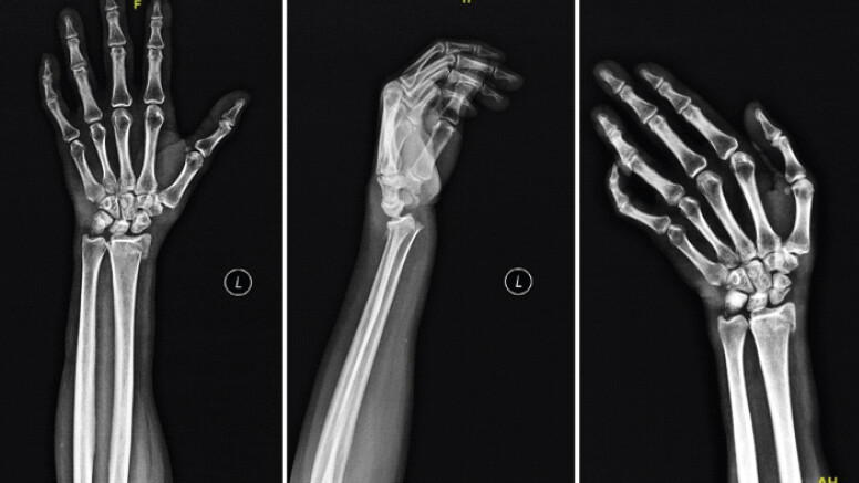
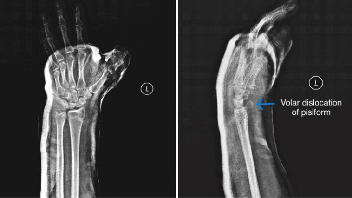
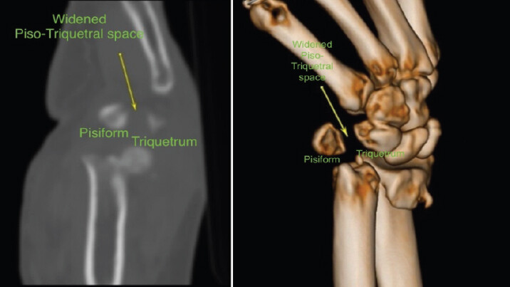
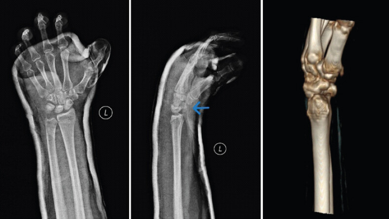
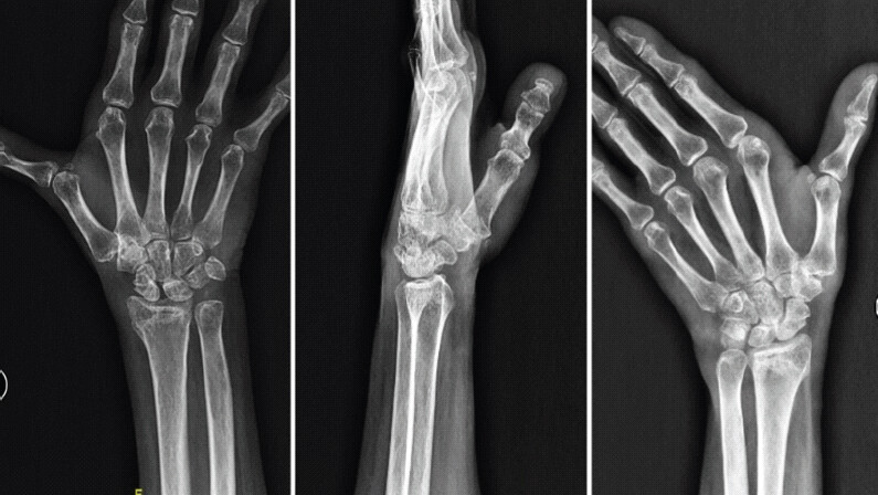
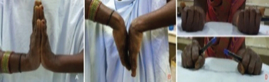
A right-hand dominant female in her early 50s presented to our emergency department following a high-velocity fall onto her outstretched left hand while she was a pillion rider on a motorcycle. She had severe pain and swelling around her left wrist. She was unable to move her wrist and had numbness over the little finger on the injured side. On examination, she had decreased sensation over her little finger and the medial half of the ring finger with no weakness of the intrinsic muscles of the hand. Standard anteroposterior, lateral and scaphoid view plain radiographs (Fig. 1) revealed an extra-articular fracture of the distal radius and a scaphoid fracture. The distal radius fracture was managed by closed reduction using a hematoma block. Post-procedure radiographs revealed a dislocated pisiform (Fig. 2). A computed tomography (CT) scan was performed; sagittal sections demonstrated volar migration of the pisiform, with the bone displaced volarly relative to its normal articulation with the triquetrum. The pisotriquetral joint space was markedly widened, and the normal congruent articular relationship was completely lost, confirming a volar dislocation pattern (Fig. 3). Subsequently, the pisiform was reduced with the wrist in palmar flexion, thereby relaxing the FCU tendon. A short below-elbow plaster cast was applied, extending to the thumb, immobilizing the scaphoid fracture. Subsequent radiographs (Fig. 4) confirmed good reduction of the fractures and the pisiform dislocation. The numbness over the little finger improved significantly within a few hours following the reduction. The cast was continued for 6 weeks when the radiographs revealed fracture union and maintenance of the pisiform position (Fig. 5). A structured physiotherapy program was started, and nine months following the injury, the patient recovered with a good range of motion in the fingers and wrist, and she managed to do her regular activities without any difficulty (Fig. 6).
Discussion
The pisiform is a sesamoid bone embedded within the tendon of the FCU and plays a crucial role in wrist biomechanics. It serves as an attachment point for several ligaments and tendons, including the pisohamate and pisometacarpal ligaments, the ulnar collateral ligament, and the transverse carpal ligament. These robust soft-tissue connections, along with its anatomical positioning, confer stability to the pisiform, making dislocations exceedingly rare [1, 2, 3].
The biomechanical mechanism underlying the concurrent distal radius fracture, scaphoid fracture, and pisiform dislocation in this case deserves systematic analysis, as each component has a classically distinct injury mechanism. Distal radius fractures typically result from axial loading through the radiocarpal joint during wrist dorsiflexion, while scaphoid fractures occur from dorsiflexion-compression forces concentrated at the scaphoid waist. Pisiform dislocation, in contrast, is driven by a fundamentally different force vector. The pisiform occupies a volar-ulnar position within the wrist, embedded in the FCU tendon and stabilized by the pisohamate and pisometacarpal ligaments. Its dislocation requires either a direct, high-energy blow to the hypothenar eminence or a sudden, violent contraction of the FCU – both generating a volar and ulnar force that, when of sufficient magnitude, overcomes the ligamentous restraints and drives the pisiform ulnarly and volarly away from its articulation with the triquetrum, as confirmed on sagittal CT in the present case. In a high-velocity pillion rider accident, the wrist is subjected to a complex, multidirectional force pattern. At the moment of impact, the outstretched hand absorbs a predominantly dorsal axial load – accounting for the distal radius and scaphoid fractures – while simultaneously, the hypothenar eminence sustains a direct volar-ulnar impact against the vehicle or road surface. This direct blow, combined with a sudden eccentric tensile load through the FCU as the wrist is forced into dorsiflexion, creates the volar shear and ulnar traction forces necessary to disrupt the pisotriquetral joint. The net result is volar and ulnar displacement of the pisiform – precisely the pattern observed on sagittal CT in this case – explaining the co-existence of injuries that would, in isolation, require entirely different mechanisms. Wrist position at the time of impact is also critical: dorsiflexion and radial deviation increase the tension within the FCU tendon and place the pisotriquetral joint under maximal distraction stress, lowering the threshold for ligamentous failure [1, 2, 3, 4, 5].
In the study by Vasilas et al., 12 out of 40 cases of distal end radius fractures had a pisiform subluxation, but none had a complete dislocation [4]. Similarly, Tajima et al., in their CT study of distal end radius fractures, found 29% pisiform malalignment, classified into three types: wide type, non-parallel type, and overriding type [5]. The volar dislocation pattern observed in the present case represents the most severe end of this spectrum, with complete loss of pisotriquetral congruity rather than subluxation.
Imaging plays a critical role in diagnosing pisiform dislocations, which may be easily missed on standard wrist radiographs if not specifically sought. On the lateral radiograph, the pisiform normally projects as an oval density overlying the palmar aspect of the triquetrum. Volar displacement results in the pisiform appearing more palmarly positioned than normal, disrupting this relationship. CT imaging is the modality of choice for characterizing the direction and degree of pisiform displacement. In the present case, sagittal sections clearly demonstrated volar migration of the pisiform relative to the triquetrum, with complete loss of articular congruity and marked joint space widening. This volar displacement pattern is mechanistically consistent with the hypothenar impact and FCU traction mechanism described above, as the volar force vector drives the pisiform away from the triquetrum in the palmar and ulnar direction. Systematic description of the displacement direction on CT – whether volar, dorsal, radial, or ulnar – should be reported in all future cases to facilitate classification and guide reduction maneuvers, since the direction of displacement logically dictates the corrective force required during closed reduction.
The proximity of the pisiform to the ulnar nerve within Guyon’s canal means that dislocation of the pisiform can lead to ulnar nerve compression, resulting in neuropraxia [6]. The volar and ulnar displacement of the pisiform observed in this case is particularly relevant to the mechanism of nerve injury. Guyon’s canal is bounded volarly by the palmar carpal ligament and dorsally by the flexor retinaculum and carpal bones, with the pisiform forming its ulnar wall. Volar migration of the pisiform reduces the cross-sectional area of the canal at Zone 1 – the proximal segment proximal to the bifurcation of the ulnar nerve – compressing the nerve before it divides into its deep motor and superficial sensory branches. The clinical picture of pure sensory deficit in the ring and little fingers with complete preservation of intrinsic muscle function is consistent with a partial Zone 1 lesion selectively affecting the superficial sensory fibers, while sparing the deep motor branch. The rapid and complete resolution of sensory symptoms within hours of pisiform reduction strongly supports a neuropraxia – a transient conduction block without axonal disruption – caused by direct mechanical compression rather than ischemia or traction.
Management of pisiform dislocations varies depending on the presence of associated injuries and the severity of the dislocation. Closed reduction and immobilization are often successful, especially when performed promptly. However, in cases where closed reduction fails or if there is persistent instability or recurrent dislocation, surgical intervention, including open reduction or pisiform excision, may be necessary. As highlighted by Letsch and de Boer, surgical exploration should be considered in cases of irreducibility or instability to address soft-tissue interposition and restore pisotriquetral congruity [11]. In cases of delayed diagnosis, capsular disruption and fibrosis may make reduction technically difficult, thereby favoring excision for pain relief and functional recovery. When the pisohamate and pisometacarpal ligaments are disrupted, surgical reconstruction may be required to restore FCU function and maintain wrist stability [7, 8, 9, 10, 11].
In the present case, the decision to pursue conservative management was based on several objective criteria indicating stability. First, closed reduction was achieved with minimal force by placing the wrist in palmar flexion – the maneuver that directly relaxes FCU tension and reverses the volar traction force responsible for the dislocation – and the pisiform relocated anatomically without requiring repeated attempts, a finding inconsistent with significant soft-tissue interposition or irreducibility. Second, post-reduction imaging confirmed restoration of pisotriquetral joint congruity with no residual widening on follow-up radiographs. Third, no clinical signs of pisotriquetral instability were elicited on stress examination. Fourth, the rapid and progressive resolution of ulnar nerve symptoms within hours of reduction confirmed decompression of Guyon’s canal, negating the need for surgical exploration. Fifth, both the distal radius and scaphoid fractures were manageable conservatively without displacement, removing any surgical indication that might have prompted simultaneous wrist exploration. Taken together, these five criteria clearly supported a non-operative approach, and the nine-month clinical and radiological outcome validated this decision.
This case underscores the importance of a high index of suspicion for associated carpal injuries, including pisiform dislocation, in patients presenting with distal radius fractures, especially when there are neurological symptoms indicative of ulnar nerve involvement. Early recognition and appropriate management are crucial to prevent long-term complications and ensure optimal functional recovery.
Conclusion
Clinicians should be vigilant about diagnosing rare concomitant injuries like pisiform dislocations with distal radius fractures. The principal teaching points from this case are threefold: first, high-velocity wrist trauma can simultaneously generate mechanistically distinct injuries – axial loading producing the distal radius and scaphoid fractures, while a direct volar-ulnar hypothenar impact combined with eccentric FCU traction produces volar and ulnar pisiform dislocation; second, sagittal CT is indispensable and should systematically characterize the direction of pisiform displacement, as this directly informs the biomechanical understanding and guides the reduction maneuver; third, volar migration of the pisiform compresses the ulnar nerve within Guyon’s canal at Zone 1, producing a partial neuropraxia that resolves reliably and promptly following anatomic reduction, without the need for surgical decompression when stability criteria are met. Prompt recognition and appropriate management typically yield excellent clinical outcomes.
Clinical Message
High Index of Suspicion: Maintain vigilance for associated carpal injuries, particularly pisiform dislocation, in patients presenting with distal radius fractures after high-velocity trauma, especially when ulnar neurological symptoms are present.
Biomechanical Awareness: Pisiform dislocation co-existing with distal radius and scaphoid fractures implies a multidirectional force mechanism – axial loading combined with a volar-ulnar hypothenar impact and FCU eccentric traction. Understanding this mechanism explains the direction of displacement and guides closed reduction.
Role of Advanced Imaging: CT is the imaging modality of choice. Reports should specify the direction of pisiform displacement – volar, ulnar, dorsal, or radial – as this has direct mechanistic and therapeutic implications.
Ulnar Nerve Localisation: Volar pisiform displacement compresses the ulnar nerve within Guyon’s canal. The clinical pattern of pure sensory deficit with preserved motor function localizes the lesion to Zone 1 (partial), and rapid resolution after reduction confirms neuropraxia rather than axonotmesis.
Non-operative Management Viability: Closed reduction with wrist in palmar flexion, confirmed anatomic restoration on imaging, absence of instability, and prompt neurological recovery collectively justify conservative management without surgical exploration.
Early Intervention: Prompt recognition and intervention significantly improve prognosis, particularly regarding neurological recovery in Guyon’s canal compression.
Conflict of Interest:
Source of Support:
Nil
Consent:
The authors confirm that informed consent was obtained from the patient for publication of this article
References
- Lall A, Shephard N, Greenbaum S, Doerre T, Wilson S, Kulick RG.Isolated carpal dislocation of the pisiform with distal radius fracture in two adults: A rare entity. J Hand Microsurg 2020;12:215-8. Google Scholar | PubMed
- Pevny T, Rayan GM, Egle D.Ligamentous and tendinous support of the pisiform, anatomic and biomechanical study. J Hand Surg Am 1995;20:299-304. Google Scholar | PubMed
- Rajeev A, Jabbar F, Chakravarthy J, Newby M.Isolated acute pisiform dislocation presenting atypically as wrist sprain in the emergency department. J Surg Case Rep 2019;2019:rjz183. Google Scholar | PubMed
- Vasilas A, Grieco V, Bartone NF.Roentgen aspects of injuries to the pisiform bone and pisotriquetral joint. J Bone Joint Surg Am 1960;42:1317-28. Google Scholar | PubMed
- Tajima T, Zenke Y, Yamanaka Y, Menuki K, Sakai A.Pisiform malalignment associated with distal radius fractures. J Orthop Sci 2018;23:511-5. Google Scholar | PubMed
- Smith JT, Keeve JP, Bertin KC, Mann RJ.Simultaneous fractures of the distal radius and scaphoid. J Trauma 1988;28:676-9. Google Scholar | PubMed
- Sharma S, Massraf A.Ulnar nerve compression as a consequence of isolated pisiform dislocation. Injury Extra 2005;36:79-81. Google Scholar | PubMed
- Letsch MT, De Boer HL, Nguyen DT.Open reduction of the dislocated pisiform bone. J Hand Microsurg 2016;8:183-4. Google Scholar | PubMed
- Petrou IG, Savioz-Leissing C, Gray A.Traumatic dislocation of the pisiform bone. J Hand Microsurg 2018;10:37-40. Google Scholar | PubMed
- Dias R, Alves-Da-Silva T, Martinho C, Guerra-Pinto F.Isolated distal pisiform dislocation: Case report. Rev Bras Ortop (Sao Paulo) 2021;57:341-4. Google Scholar | PubMed
- Saglam F, Dagtas MZ, Sağlam S, Baysal O.Locked traumatic pisiform dislocation: A case report. JBJS Case Connect 2019;9:e0278. Google Scholar | PubMed
- Mahmood F, Mehta M, Kakkar R.Pisiform dislocation. BMJ Case Rep 2021;14:e237482. Google Scholar | PubMed
© 2026 Journal of Orthopaedic Case Reports - Published by Indian Orthopaedic Research Group
About the Authors
-

Dr. Prabhu Muthiahpandian
-

Dr. Pasupathy Palaniappan
-

Dr. Sanjey Sivakumar
-

Dr. Karthikeyan Dhandapani
-

Dr. Raja Vigneswar
-

Dr. Rahul Kokkiligadda
Related Articles in Journal of Orthopaedic Case Reports
 September 1, 2026 Proximal Row Carpectomy for Established Post-Traumatic Radiocarpal Arthritis Following Distal Radius Volar Rim and Scaphoid Fractures: A Case Report
 September 1, 2026 Concomitant Distal Radius Fracture with Scaphoid Fracture-Dislocation and Carpal Instability: A Case Report
 August 1, 2026 Volar Carpometacarpal Dislocation Associated with Fracture of the Second, Third, and Fourth Metacarpal Bases Treated by Closed Reduction and Percutaneous Fixation: A Case Report
 June 1, 2026 Nonvascularized Iliac Crest Autograft for Wrist and Hand Reconstructions: A Report of Two Clinical Cases and Literature Review