


Introduction
Ewing sarcoma is an aggressive malignancy characterized by the rapid proliferation of small, round blue cells. It commonly metastasizes early to the lungs, bones, and lymph nodes, leading to a poor prognosis. It is described as the second most common tumor after osteosarcoma, mostly affecting 5–20-year-olds with male predominance, usually involving the long bones of the extremities, such as the femur 16.4%, the pelvis, and the ribs [1,2].
Ewing sarcoma family of tumors (ESFTs) consists of extraosseous Ewing sarcoma, skeletal Ewing sarcoma, primitive neuroectodermal tumor, and Askin tumor.
Askin tumor is a rare malignant neoplasm of neuroectodermal origin, which develops from the soft tissues of the thoracopulmonary wall; it is predominantly seen in children, most commonly in males. The patients may have dyspnea, cough, Horner’s syndrome, and regional lymphadenopathy. They usually present on computed tomography (CT) with a large, heterogeneous, extrapulmonary mass lesion with or without destruction of the ribs and frequently associated with pleural effusion. 10% of the patients usually have evidence of metastasis at diagnosis. Common sites of metastasis are the lungs, bone marrow, bone, and mediastinal lymph nodes. The treatment consists of neoadjuvant chemotherapy followed by surgical excision with or without adjuvant radiotherapy [3, 4]. It is associated with poor prognosis and short survival because local recurrence and metastases are frequently seen in Askin tumor. The reported overall survival is approximately 60% at 5 years [3, 5]. In an attempt to further define prognostic factors, appropriate management and treatment outcome of this rare neoplasm, we reviewed our experience with this relatively rare round cell tumor. Here, we report a case of extraosseous Ewing sarcoma with Askin tumor in a 12-year-old female.
Case Report
We present a case of a 12-year-old white Middle Eastern female with no significant past medical history who presented to the outpatient clinic complaining of right thigh pain and swelling associated with limping for 4 months. With normal regular pulses and normal vital signs. She has no family history of Ewing sarcoma or Askin tumor, and she denied any history of recent trauma, prolonged immobilization, or infection. Furthermore, denying any history of back pain, chest pain, chest tightness or discomfort, no fever, night sweats, or weight loss.
A timeline, including symptom onset, diagnostic steps, treatment interventions, and follow-up, is provided in Table 1.
Timeline of Clinical Events
| Event | Timeline |
|---|---|
| Symptom onset | 4 months before admission |
| Initial Imaging (X-ray, MRI, CT) | At admission |
| Biopsies performed | Shortly after imaging |
| Diagnosis confirmed | Post-biopsy |
| Start of chemotherapy | After diagnosis (VDC protocol) |
| Surgical resection | Post-chemotherapy |
| Post-op chemo/radiotherapy | Following surgery |
| Bone marrow biopsy | Post-treatment |
On examination, it revealed an immobile, tender, tense swelling in the right thigh without any local increase in temperature. The range of motion of the joint and distal neurovascular status were normal. No palpable inguinal lymph nodes.
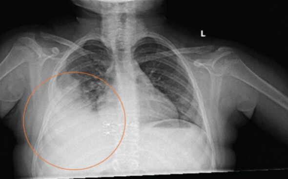
On investigation, as shown in Table 2, white blood cells, erythrocyte sedimentation rate, and C-reactive protein were elevated. Fig. 1 revealed that a chest X-ray was done on the day of admission as a clinical protocol of the hospital.
Laboratory Results upon Admission Day
| Test Name | Normal Range (children) | Results |
|---|---|---|
| White blood cells (WBCs) | 4.6-10.2mg/dl | 19.6 |
| Neutrophils | 2-6.9 mg/dl | 15.5 |
| Erythrocyte sedimentation rate (ESR) | 0-20 g/dl | 81 |
| C-Reactive Protein (CRP) | 8-10 mg/L | 257 |
| Calcium | 86-10.2mg/dl | 9.5 |
| Alkaline phosphatase (ALP) | 40-480unit/L | 380.9 |
A right thigh X-ray was done, which revealed that there was about 7.5 × 5 × 5 soft-tissue lesion, also confirming a lytic bone lesion. Fig. 2 shows magnetic resonance imaging findings revealing a heterogeneous intramedullary mass lesion of the right femoral shaft with a soft-tissue component extending over a distance of 21cm in length, which appears hypointense on T1 and hyperintense on T2-weighted sequence with heterogeneous post-contrast enhancement and heavy diffusion restriction, which was associated with diffuse cortical bone destruction and large soft-tissue extraosseous extension surrounding the affected bone. Similar enhanced abnormal intramedullary signals were seen in the left iliac bone and distal right femoral metaphysis, with the largest measures up to 17 × 16 mm in the left iliac bone. Suggestive of Ewing sarcoma with bony metastasis.
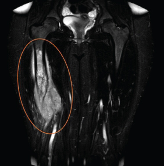
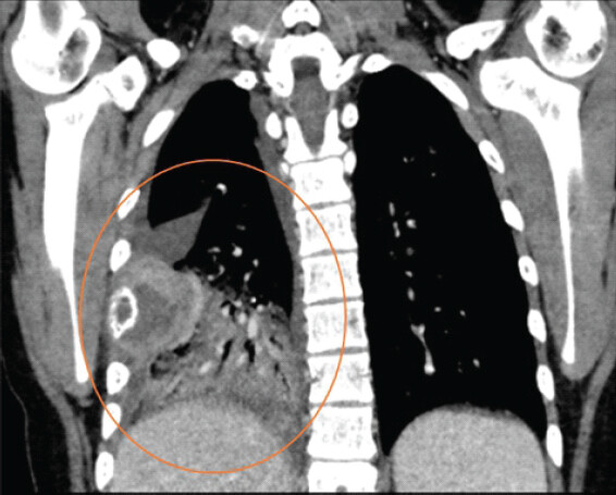
Fig. 3 shows a CT scan with contrast for lung, mediastinal and bone were done which presented bilateral lower lung lobe nodules: The largest on the right measures 2 cm and on the left measures 1.5 cm and permeative destructive lesion in the right 5th rib associated with large soft tissue component measuring 9 cm and subsequent consolidation of the right lower lobe, with rim of right sided pleural effusion, which raises suspicion for Askin tumor, also a lytic bone lesion in the iliac side of the left sacroiliac joint measuring 2 cm.
Under sedation and ultrasound guidance, a needle biopsy was performed on the right thigh and posterior chest wall. Histopathological examination revealed microscopically small round neoplastic cells with hyperchromatic nuclei and minimal to focally moderate vacuolated cytoplasm. Mitotic activity was observed. Areas of tumor necrosis are seen; the background shows a desmoplastic reaction.
On immunohistochemistry, the tumor cells were positive for CD99 (membranous), whereas desmin, myogenin, CD45, and SOX10 were negative. A final diagnosis of Ewing sarcoma of the right thigh with bone metastases and Askin tumor.
Based on the patient’s history, examination findings, and previous investigation, she was scheduled for pre-operative chemotherapy management, as a vincristine, doxorubicin, cyclophosphamide protocol was done over 6 cycles of chemotherapy. Re-evaluation chest CT and X-ray demonstrated a significant decrease in the previously described right 5th rib destructive lesion with residual soft-tissue component. Subsequently, the patient underwent a radical resection of the femur tumor as follows: an intra-operative procedure. After segmental bone resection, it underwent extracorporeal radiation; fixation and reaming were done for the proximal intramedullary canal and distal intramedullary canal with preservation of the growth plate area, then the reduction and fixation of bone were checked under C-arm image; the procedure was successfully completed. Post-operative chemoradiation was done. Bone marrow biopsy and aspiration were repeated and confirmed a normocellular marrow with trilineage hematopoiesis to ensure that the patient was in good health.
Discussion
Ewing sarcoma is a highly metastatic form of sarcoma that ranks as the second most prevalent primary malignant bone tumor, constituting 10–15% of all soft bone sarcomas, predominantly afflicting males between 10 and 15 years of age. Extraosseous Ewing sarcoma and Askin tumors are types of ESFT that are characterized by the presence of non-random chromosomal translocation producing fusion genes 11; 22-q24 [2]. Ewing sarcoma classically presents with leg pain, stiffness, or swelling; more than 50% of patients with Ewing sarcoma have intermittent pain that worsens at night. Around 20% of patients present with metastatic disease at the time of diagnosis, and among these cases more than 20% have lung or pleural involvement [6].
Askin tumors maintain a low incidence regardless of the new diagnostic technique. They are more common in males, and 80% of patients are children and young adults; however, there are reported cases of elderly patients. Usually, patients come with ambiguous symptoms including a palpable mass in the thorax, either painful or painless (the most common finding), pleuritic pain, dyspnea, fever, cough, and weight loss [3, 4].
Our 12-year-old female patient presented with extraosseous Ewing sarcoma (Ewing sarcoma) with bony metastases and an Askin tumor at the time of diagnosis, with an unusual presentation of limping with mild swelling in the right thigh, pain and tenderness, but she denied any fever, night sweats, weight loss, or chest symptoms. During the investigation, we incidentally discovered two masses in the lung with minimal right-sided pleural effusion. This is different from typical presentations of Askin tumor, which often manifest with respiratory symptoms such as dyspnea and chest pain. The lack of thoracic symptoms in our patient underscores the importance of comprehensive imaging in cases of unexplained limb pain and swelling.
The presentation of Ewing sarcoma with bony metastasis as an Askin tumor is extremely rare. Almost all literature reviews reported each of these types separately, with only 17 cases identified in the literature reviewing Askin tumor. Table 3 presents a summary of selected reported cases, their treatment approaches, and outcomes. After an extensive review of the literature, we have not found any study reporting Ewing sarcoma of bone metastasis with Askin tumor at the time of diagnosis. This case uniquely shows their co-occurrence, a combination that has not been previously detailed in the literature.
Selected reported Askin Tumor Cases in Literature
| Study | Treatment Approach | Survival Outcome |
|---|---|---|
| Zhang et al. (2016) | Surgery + Chemotherapy | ~60% 5-year OS |
| Laskar et al. (2011) | Neoadjuvant chemo, surgery, radiation | Variable, high recurrence |
| Singh et al. (2016) | Chemo + Radiotherapy | Poor prognosis, rapid recurrence |
The co-occurrence of Ewing sarcoma with Askin tumor, while extraordinarily rare, can be better understood by examining their shared histogenetic and molecular characteristics. Both are part of the ESFTs, defined by the presence of specific chromosomal translocations, most commonly t(11;22)(q24;q12), resulting in the EWSR1-FLI1 fusion gene. This fusion gene acts as an aberrant transcription factor that drives oncogenesis by dysregulating gene expression involved in cell proliferation, differentiation, and apoptosis. The identification of this translocation in both intraosseous and thoracopulmonary tumors supports the hypothesis of a shared origin from primitive neuroectodermal cells, albeit arising in different anatomical locations.
From a radiological perspective, positron emission tomography-CT (PET-CT) has become an essential tool in the staging and restaging of Ewing sarcoma and related tumors. It provides valuable information on metabolic activity, enabling detection of both skeletal and soft-tissue involvement with high sensitivity. In this case, although PET-CT was not initially used, its incorporation in follow-up evaluations could provide superior assessment of treatment response and early detection of recurrence.
Histologically, both tumors consist of small, round blue cells. Immunohistochemistry plays a critical role in differentiating them from other small round cell tumors. In our case, tumor cells showed diffuse membranous positivity for CD99, which, although not specific, is highly sensitive for ESFT. The negative markers (desmin, myogenin, CD45, SOX10) helped exclude rhabdomyosarcoma, lymphoma, and other mimics. In addition, newer markers such as NKX2.2, FLI1, and molecular confirmation by fluorescence in situ hybridization or reverse transcription-polymerase chain reaction for EWSR1 rearrangement can enhance diagnostic accuracy, especially in ambiguous cases [7].
The other differential diagnoses are inflammatory disorders and other malignancies. Clinical symptoms of Askin tumor mimic inflammatory disorders such as osteomyelitis, empyema, and TB, all of which can present with bone pain and systemic symptoms. On the side of malignancies in children include lymphoma (lymphadenopathy, splenomegaly), osteosarcoma, and neuroblastoma (elevated urinary catecholamines) [3, 5, 8]. A comparative overview of these conditions is presented in Table 4, highlighting their distinguishing clinical features and diagnostic modalities.
Differential Diagnosis Comparison
| Condition | Distinguishing Features | Diagnostic Tools |
|---|---|---|
| Ewing Sarcoma | Small round blue cell tumor; CD99+, EWSR1 translocation | Histology, IHC, FISH/PCR |
| Askin Tumor | Chest wall origin, same histologic features as ES | Imaging, location, IHC |
| Osteomyelitis | Infection markers, fever, elevated CRP | Blood tests, imaging |
| Lymphoma | Lymphadenopathy, systemic symptoms | Biopsy, immunophenotyping |
| Neuroblastoma | Adrenal origin, elevated catecholamines | Urine catecholamines, MIBG scan |
Given the aggressive nature and high metastatic potential of both tumors, a multimodal approach involving neoadjuvant chemotherapy, surgical resection, and adjuvant radiotherapy remains the cornerstone of treatment. Future therapeutic directions may include EWS-FLI1-targeted therapies and immune checkpoint inhibitors, which are currently under investigation in clinical trials.
Due to the disease’s rarity, there are not many small-scale, single-institution research studies and no established treatment guidelines. Emerging therapies, such as proton therapy, offer targeted approaches to minimize damage to surrounding healthy tissue, especially in pediatric cases. Furthermore, clinical trials exploring checkpoint inhibitors and molecular inhibitors for EWS-FLI1 fusion proteins may provide new hope for aggressive and metastatic disease management [9, 10]. The successful management of this case underscores the importance of a multidisciplinary approach, integrating oncology, surgery, and radiology expertise. Individualized treatment plans tailored to pediatric patients’ unique needs can significantly improve survival rates. This plays a major role when combined with early clinical suspicion, especially in resource-limited settings, to overcome the challenging diagnoses of rare tumors such as Askin tumor and metastatic Ewing sarcoma due to limited access to advanced imaging and genetic studies.
Beyond medical management, addressing the psychosocial impact of such a diagnosis is critical, particularly in pediatric patients. Comprehensive care should include psychological support, rehabilitation, and counseling for both the patient and their family to improve overall quality of life. Furthermore, detecting a disease in its early stages often allows us an improved prognosis, reducing complications and engaging the patient with daily activities. This also reduces the need for more intensive and expensive interventions later. Specialists from various fields collaborate to develop a personalized treatment strategy that optimizes outcomes.
This case reinforces the need for heightened clinical suspicion, especially when initial symptoms are non-specific, and advocates for integrating advanced imaging and molecular diagnostics in early workup to facilitate timely and precise management [1,11].
Conclusion
This case highlights the rare co-occurrence of Askin tumor and metastatic Ewing sarcoma at initial diagnosis in a pediatric patient. Early diagnosis, comprehensive imaging, and a multidisciplinary approach are critical in managing such complex presentations. Future studies should explore the molecular relationship between these entities to inform targeted therapies and improve outcomes.
Askin tumor, also called Ewing sarcoma of the chest wall, is an extremely rare disease and is often misdiagnosed. Hence, careful monitoring should be prioritized. Appropriate and early multimodal treatment plans should be established during diagnosis to achieve satisfactory outcomes and to point out the significance of careful and early evaluation, even for typical clinical signs. This case highlights the importance of considering early diagnosis for intervention to occur quickly and to obtain a favorable outcome and optimal survival for the patient. Hence, the physicians should maintain a high index of suspicion when encountering patients, particularly children and young adults. It also emphasizes the importance of a multidisciplinary approach to ensure accurate diagnosis, effective treatment, and improved outcomes.
Clinical Message
Unexplained limb pain in children warrants thorough evaluation for metastatic Ewing sarcoma, including asymptomatic thoracopulmonary involvement, to facilitate early diagnosis and multidisciplinary treatment.
Conflict of Interest:
Nil
Source of Support:
Nil
Consent:
The authors confirm that informed consent was obtained from the patient for publication of this article
References
- Thomas A, Obeidat N, Darweesh M. Thoracic Ewing's sarcoma: A case report. Cureus 2022;14:e24150. Google Scholar | PubMed
- Fedeli MA, Marras V, Fara AM, Deiana A, Lobrano R, Cossu A. Primary Ewing sarcoma of the lung: A systematic review of the recent literature. Ann Diagn Pathol 2023;65:152152. Google Scholar | PubMed
- Laskar S, Nair C, Mallik S, Bahl G, Pai S, Shet T. Prognostic factors and outcome in Askin-Rosai tumor: A review of 104 patients. Int J Radiat Oncol Biol Phys 2011;79:202-7. Google Scholar | PubMed
- Zhang KE, Lu R, Zhang P, Shen S, Li X. Askin's tumor: 11 cases and a review of the literature. Oncol Lett 2016;11:253-6. Google Scholar | PubMed
- Singh A, Abhinay A, Kumar A, Prasad R, Ghosh A, Mishra OP. Askin tumor: A rare neoplasm of thoracopulmonary region. Lung India 2016;33:196-8. Google Scholar | PubMed
- Seren Durer DP. Ewing Sarcoma: Treatment and Management: Point of Care. StatPearls; 2024. Available from: https://www.statpearls.com/point-of-care/21419. [Last accessed 08 August 2026]. Google Scholar | PubMed
- Ozaki T. Diagnosis and treatment of Ewing sarcoma of the bone: A review article. J Orthop Sci 2015;20:250-63. Google Scholar | PubMed
- Xu Q, Xu K, Yang C, Zhang X, Meng Y, Quan Q. Askin tumor: Four case reports and a review of the literature. Cancer Imaging 2011;11:184-8. Google Scholar | PubMed
- Setty BA, Gikandi A, DuBois SG. Ewing sarcoma drug therapy: Current standard of care and emerging agents. Paediatr Drugs 2023;25:389-97. Google Scholar | PubMed
- Strauss SJ, Berlanga P, McCabe MG. Emerging therapies in Ewing sarcoma. Curr Opin Oncol 2024;36:297-304. Google Scholar | PubMed
- Zhang S, Li Y, Wang R, Song B. Ewing's sarcoma/primitive neuroectodermal tumor of the kidney: A case report and literature review. Transl Androl Urol 2019;8:562-6. Google Scholar | PubMed
Related Articles in Journal of Orthopaedic Case Reports
 September 1, 2026 Iliopsoas Abscess as the Initial Manifestation of Periprosthetic Hip Joint Infection after Total Hip Arthroplasty: A Case Report and Literature Review
 September 1, 2026 Limb Salvage for Entire Tibial Ewing Sarcoma in a 4-year-Old Child Using Extracorporeal Irradiation and Free Flap Reconstruction: A Case Report and Surgical Technique
 September 1, 2026 Angular Improvement with Bracing Over 7 Years in Anterolateral Tibial Bowing without Neurofibromatosis Type 1: A Case Report
 September 1, 2026 Clinical Outcomes of Staged Bilateral Middle Trapezius Transfer for Irreparable Supraspinatus Tendon Tears: A Case Report