


Introduction
Degenerative joint disease of the knee is a premier global contributor to chronic musculoskeletal disability and persistent pain. In India, community epidemiological surveys indicate that symptomatic knee osteoarthritis (OA) affects between 22% and 39% of individuals above 40 years of age, fueled by an expanding elderly population and cultural activities demanding repetitive deep flexion, squatting, and cross-legged sitting. Within this clinical spectrum, degenerative meniscal tears are increasingly understood not as isolated age-related radiographic artifacts but as active drivers of the osteoarthritic process.[1]
The menisci are fibrocartilaginous crescents engineered with circumferentially oriented collagen bundles and radially aligned tie fibers designed to distribute approximately 60% of joint compressive loads, dampen axial shock, maintain joint congruity, and lubricate articular surfaces. With senescent matrix degeneration and repeated mechanical strain, loss of proteoglycan matrix and collagen fragmentation impair the ability to withstand circumferential hoop tension. This leads to meniscal extrusion, uncontained contact pressure on articular cartilage, accelerated chondrocyte apoptosis, and synovial inflammation.[2]
Standard non-operative measures – such as oral non-steroidal anti-inflammatory drugs (NSAIDs), physical therapy, and intra-articular corticosteroids or hyaluronic acid – offer temporary symptom relief but fail to promote tissue repair or reverse structural cartilage and meniscal deterioration. Conversely, arthroscopic meniscectomy in degenerative tears often accelerates osteoarthritic progression by permanently increasing focal joint contact stress. Consequently, autologous orthobiologics have emerged as an appealing paradigm for biological joint preservation.
Platelet-rich plasma (PRP) contains concentrated autologous platelets that secrete bioactive growth factors upon activation – notably platelet-derived growth factor (PDGF), transforming growth factor-beta (TGF-β), and vascular endothelial growth factor (VEGF) – facilitating fibrocartilage healing and dampening synovial inflammation. However, conventional PRP contains variable quantities of cellular elements and leukocytes, which may provoke inflammatory synovitis and clinical flares. Growth factor concentrate (GFC) is an advanced orthobiologic formulation derived from autologous platelets activated in vitro to harvest a purified, acellular cytokine pool without leukocytes or cell fragments. Despite promising biological attributes, direct comparative clinical trials evaluating PRP versus GFC in early degenerative meniscal lesions are limited. Therefore, this randomized controlled trial was conducted to compare the functional and radiological outcomes of intra-articular PRP versus GFC in patients with degenerative meniscal lesions in Kellgren–Lawrence (K-L) Grade I and II knee OA.
Materials and Methods
Study design and ethical approval
This prospective, single-center, hospital-based, randomized controlled trial was conducted in the Department of Orthopaedics at a tertiary care university hospital over a period of 18 months. Ethical clearance was granted by the Institutional Ethics Committee (Ref. No. TMU IEC/PG 2024-25/128; Dated June 10, 2024) in accordance with the Declaration of Helsinki and Indian Council of Medical Research guidelines. Written informed consent was obtained from all patients before study participation.
Patient selection and eligibility criteria
Adult patients presenting to the Orthopaedics Outpatient Department with persistent knee pain and functional limitations were prospectively screened. Inclusion criteria were (1) age between 18 and 65 years, (2) symptomatic knee OA diagnosed as K-L Grade I or II on standard weight-bearing anteroposterior and lateral radiographs, and (3) magnetic resonance imaging (MRI)-documented degenerative meniscal lesions (Stoller Grade I, II, or stable Grade III tear). Exclusion criteria were acute knee trauma within the preceding 12 months; prior intra-articular injections (steroids, viscosupplementation, or orthobiologics) within the past year; advanced OA (K-L Grade III or IV); active inflammatory arthropathy, joint infection, systemic malignancy, coagulopathies, or uncontrolled diabetes mellitus; baseline platelet count <1.5 × 10⁵/µL; and multi-joint involvement.
Randomization and interventions
Eligible participants were randomized into three parallel groups (1:1:1 allocation ratio) using computer-generated random numbers (n = 18/group):
-
Group A (PRP group): Received two intra-articular injections of autologous PRP administered 4 weeks apart
-
Group B (GFC group): Received two intra-articular injections of autologous GFC administered 4 weeks apart
-
Group C (Control group): Received two intra-articular injections of sterile Normal Saline (0.9% NaCl) administered 4 weeks apart.
Biologic preparation and administration protocol
PRP preparation
Under strict aseptic precautions, 28–42.5 mL of autologous venous blood was drawn into acid citrate dextrose tubes. A two-stage centrifugation protocol was utilized: An initial soft spin at 3,000 rpm for 3 min to separate red blood cells, followed by a hard spin of the plasma fraction at 4,000 rpm for 15 min to isolate the platelet pellet, yielding approximately 3 mL of buffy-coat-poor PRP.
GFC preparation
Eight milliliters of autologous venous blood was collected into dedicated GFC vacutainers without chemical anticoagulants. Blood was incubated for 30 min at room temperature to allow physiologic activation of platelets and release of alpha-granule growth factors. Centrifugation was then performed at 3,400 rpm for 10 min, yielding approximately 3 mL of acellular, growth-factor-enriched supernatant.
Injection procedure
Patients were positioned supine with the knee flexed to 90°. No local anesthesia was administered to avoid cellular cytotoxicity. Under strict aseptic technique, 3 mL of the assigned injectate was injected into the intra-articular space through an anterolateral approach using a 22-gauge needle. The knee was actively mobilized through gentle flexion-extension cycles to disperse the injectate. Patients were monitored for 60 min. Oral tramadol (50 mg as needed, maximum 72 h) was permitted for post-injection pain; systemic NSAIDs were prohibited.
Outcome assessment and follow-up
Patients were evaluated at baseline and at 1, 3, 6, and 9-month post-injection. Pain intensity was evaluated through the 10-point Visual Analog Scale (VAS). Functional status was evaluated using the Western Ontario and McMaster Universities OA Index (WOMAC). Radiological joint and meniscal status were evaluated using 1.5-Tesla MRI scored according to the whole-organ MRI score (WORMS) meniscal scoring system at baseline and at 9 months.[3]
Statistical analysis
Statistical calculations were executed using IBM Statistical Package for the Social Sciences Statistics version 26.0. Continuous data were summarized as mean ± standard deviation and categorical variables as counts and percentages. Intergroup baseline comparisons were performed using Analysis of variance (ANOVA) and the Chi-square test. Longitudinal within-group and between-group changes were assessed using repeated-measures ANOVA followed by Tukey’s post hoc test. A P < 0.05 was defined as statistically significant.
Results
A total of 58 patients were initially randomized. Four patients were lost to follow-up, leaving 54 patients (18/group) who completed all clinical and imaging visits at 9 months and were included in the conclusive analysis. Baseline demographic and clinical characteristics are summarized in Table 1.
Baseline demographic and clinical characteristics across groups
| Parameter | Control (n=18) | GFC (n=18) | PRP (n=18) | Statistic | P-value |
|---|---|---|---|---|---|
| Age (years, mean±SD) | 58.24±10.00 | 55.63±9.10 | 57.28±11.28 | F=0.324 | 0.725 |
| Age range (Min-Max) | 45-75 | 41-74 | 42-78 | — | — |
| Gender: Male (%) | 5 (27.8) | 6 (33.3) | 6 (33.3) | χ2=0.172 | 0.918 |
| Gender: Female (%) | 13 (72.2) | 12 (66.7) | 12 (66.7) | — | — |
| K-L Grade I (%) | 2 (11.1) | 4 (22.2) | 2 (11.1) | χ2=1.118 | 0.556 |
| K-L Grade II (%) | 16 (88.9) | 14 (77.8) | 16 (88.9) | — | — |
| Baseline VAS score | 6.00±0.75 | 6.21±0.71 | 6.33±0.69 | F=0.661 | 0.521 |
| Baseline WOMAC score | 56.40±8.41 | 59.16±8.26 | 59.58±8.72 | F=0.319 | 0.728 |
| Baseline WORMS score | 10.65±1.34 | 10.58±1.41 | 10.22±1.28 | F=0.369 | 0.693 |
VAS: Visual Analog Scale, SD: Standard deviation, WORMS: Whole-organ magnetic resonance imaging score, GFC: Growth factor concentrate, PRP: Platelet-rich plasma, K-L: Kellgren-Lawrence, WOMAC: Western Ontario and McMaster Universities Osteoarthritis Index
*P < 0.05 was considered statistically significant.
Pain assessment (VAS)
Baseline VAS scores showed equivalent baseline pain across groups (Control: 6.00 ± 0.75, GFC: 6.21 ± 0.71, PRP: 6.33 ± 0.69; P = 0.521). During the 9-month follow-up, the GFC group achieved superior and sustained pain relief compared with PRP and control (Table 2). In Group B (GFC), VAS scores decreased progressively to 4.63 ± 0.88 at 3 months, 4.26 ± 0.81 at 6 months, and 3.58 ± 0.74 at 9 months (overall reduction: −2.63 points; −42.35%). In Group A (PRP), VAS scores decreased to 5.11 ± 0.77 at 9 months (reduction: −1.22 points; −19.27%). Conversely, Group C (Control) showed significant worsening of pain (+1.06 points; +17.67%; P < 0.0001). Differences between groups were statistically significant at 3, 6, and 9 months (P < 0.0001).
Comparison of VAS pain scores over 9 months
| Interval | Control (n=18) | GFC (n=18) | PRP (n=18) | F-value | P-value |
|---|---|---|---|---|---|
| Baseline | 6.00±0.75 | 6.21±0.71 | 6.33±0.69 | 0.661 | 0.5207 |
| 3 Months | 6.41±0.84 | 4.63±0.88 | 5.50±0.82 | 18.579 | <0.000001* |
| 6 Months | 6.65±0.92 | 4.26±0.81 | 5.44±0.79 | 36.132 | <0.000000001* |
| 9 Months | 7.06±0.96 | 3.58±0.74 | 5.11±0.77 | 36.819 | <0.000000001* |
| Mean change (%) | +1.06 (+17.67) | -2.63 (-42.35) | -1.22 (-19.27) | — | <0.0001* |
GFC: Growth factor concentrate, PRP: Platelet-rich plasma, VAS: Visual Analog Scale
*
P < 0.05 was considered statistically significant.
Functional assessment (WOMAC index)
Functional recovery across pain, stiffness, and physical performance domains demonstrated marked improvements in both active intervention arms (Table 3). Group B (GFC) achieved the greatest reduction in WOMAC score, falling from 59.16 ± 8.26 at baseline to 39.97 ± 6.21 at 9 months (−19.19 points; 32.44% improvement). Group A (PRP) improved from 59.58 ± 8.72 to 47.33 ± 7.05 (−12.25 points; 20.56% improvement). In contrast, Group C (Control) showed progressive functional decline, with WOMAC increasing from 56.40 ± 8.41 to 61.21 ± 9.14 (+4.81 points; +8.53% deterioration; P < 0.000001). Intergroup differences were statistically significant from 3 months onward.
Comparison of WOMAC functional scores over 9 months
| Interval | Control (n=18) | GFC (n=18) | PRP (n=18) | F-value | P-value |
|---|---|---|---|---|---|
| Baseline | 56.40±8.41 | 59.16±8.26 | 59.58±8.72 | 0.319 | 0.7283 |
| 3 Months | 59.10±8.75 | 47.67±7.54 | 48.81±7.92 | 7.029 | 0.0020* |
| 6 Months | 59.92±8.91 | 43.83±6.88 | 48.55±7.41 | 13.769 | 0.000017* |
| 9 Months | 61.21±9.14 | 39.97±6.21 | 47.33±7.05 | 20.796 | <0.000001* |
| Mean change (%) | +4.81 (+8.53) | −19.19 (−32.44) | −12.25 (−20.56) | — | <0.0001* |
GFC: Growth factor concentrate, PRP: Platelet-rich plasma, WOMAC: Western Ontario and McMaster Universities OA index
*
P < 0.05 was considered statistically significant.
Radiological outcome and structural changes (WORMS score)
WORMS evaluation demonstrated significant structural improvement and stabilization in both active biologic groups at 9 months (Table 4). Mean WORMS score decreased from 10.58 ± 1.41 to 6.11 ± 1.02 (−4.47 points; −42.25%) in the GFC group, and from 10.22 ± 1.28 to 6.11 ± 1.08 (−4.11 points; −40.22%) in the PRP group. In contrast, the control group showed minimal change (10.65 ± 1.34–10.41 ± 1.29; −0.24 points; −2.25%; P < 0.0001). Post hoc testing demonstrated no statistically significant difference in 9-month WORMS score between GFC and PRP.
Baseline and 9-month radiological WORMS score comparison
| Timeline | Control (n=18) | GFC (n=18) | PRP (n=18) | F-value | P-value |
|---|---|---|---|---|---|
| Baseline (Pre-intervention) | 10.65±1.34 | 10.58±1.41 | 10.22±1.28 | 0.369 | 0.693 |
| 9 Months (Post-intervention) | 10.41±1.29 | 6.11±1.02 | 6.11±1.08 | 48.392 | <0.000000000001* |
| Absolute change | −0.24 | −4.47 | −4.11 | — | <0.0001* |
| Percentage improvement (%) | −2.25 | −42.25 | −40.22 | — | <0.0001* |
WORMS: Whole-organ magnetic resonance imaging score, GFC: Growth factor concentrate, PRP: Platelet-rich plasma
*
P < 0.05 was considered statistically significant.
Discussion
Degenerative meniscal lesions in the setting of early knee OA (K-L Grade I and II) present a challenging clinical entity. Because menisci play a vital role in circumferential load distribution, disruption of the fibrocartilaginous network elevates tibiofemoral peak stresses, accelerating articular chondral loss. This randomized controlled trial was conducted to compare the clinical and radiological efficacy of intra-articular PRP versus GFC. The biological rationale for PRP includes the release of growth factors involved in tissue repair.[4]
The demographic profile in our study revealed a mean age of 57.28 ± 11.28 years in the PRP group and 55.63 ± 9.10 years in the GFC group, with an overall female predominance of 68.5%. This mirrors established epidemiological data, reflecting the confluence of postmenopausal hormonal withdrawal, microstructural collagen degeneration, and biomechanical stress.
A major finding of this study is the marked clinical superiority of GFC over PRP in pain alleviation (42.35% vs. 19.27% VAS reduction) and functional recovery (32.44% vs. 20.56% WOMAC improvement) at 9 months. Although PRP yielded meaningful clinical gains – concordant with meta-analyses by Liang et al. (2025) and Khalid et al. (2024) – GFC established clear statistical superiority across all follow-up intervals. The superior efficacy of GFC is grounded in molecular biology. Conventional PRP retains cellular elements, including pro-inflammatory leukocytes and neutrophils, which can release reactive oxygen species, matrix metalloproteinases, and catabolic cytokines (interleukin-1 beta, tumor necrosis factor-alpha) that induce transient post-injection synovitis. Conversely, GFC is processed through controlled in vitro platelet activation that isolates high concentrations of anabolic growth factors (PDGF, TGF-β, VEGF, EGF) in an acellular supernatant completely devoid of leukocytes. This eliminates inflammatory post-injection flares while providing sustained anabolic signaling for fibrochondrocyte viability and synovial equilibrium.[5–13] Previous studies have also evaluated PRP in MRI-confirmed degenerative meniscal lesions and meniscal injuries.[14–17].
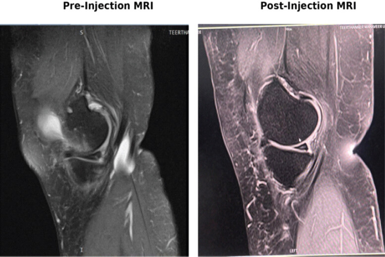
WORMS analysis showed significant structural stabilization and effusion resolution in both biologic groups at 9 months (−4.47 in GFC, −4.11 in PRP) relative to saline control (−0.24). As demonstrated in Fig. 1, follow-up MRI revealed marked resolution of joint effusion and stabilization of meniscal contours without tear extension or extrusion progression. Interestingly, while GFC was superior in subjective pain and function scores, structural scores on WORMS were statistically comparable between GFC and PRP. This observation is consistent with findings reported by Harna et al. (2024) and Varun Gopinath et al. (2024), indicating that biological suppression of synovitis, pain modulation, and joint lubrication occur rapidly, whereas macro-structural fibrocartilaginous remodeling requires extended timelines to be fully captured on MRI. The biological basis for these structural observations is also consistent with literature on growth factors in cartilage repair and meniscal structure and hoop tension.[18–20]
Limitations and future scope
This study has several limitations. First, the small sample size, with 54 participants completing the study, limits statistical power and generalizability. Second, its single-center design limits external validity. Third, the 9-month follow-up was insufficient to assess long-term durability, OA progression, or whether treatment could delay or prevent knee arthroplasty. Fourth, four participants were lost to follow-up, and analysis was limited to study completers; therefore, attrition bias cannot be excluded.
Fifth, only K-L Grade I and II OA were included, limiting applicability to moderate or advanced disease. Sixth, meniscal lesions were not stratified according to morphology, location, size, extrusion, or root involvement, resulting in potential clinical heterogeneity. Seventh, MRI was performed only at baseline and 9 months using conventional 1.5-T MRI, without advanced quantitative techniques such as T2 mapping; the sensitivity of WORMS for subtle tissue-level changes may also be limited.
Eighth, the biological composition of GFC and PRP was not quantitatively characterized, including growth-factor, platelet, and leukocyte concentrations. Ninth, blinding of participants, clinicians, and outcome/MRI assessors was not described, creating potential performance and assessment bias. Tenth, clinical outcomes were assessed mainly using VAS and WOMAC, without additional activity or quality-of-life measures. Finally, cytokine levels, inflammatory biomarkers, and long-term structural progression were not assessed; therefore, mechanistic or disease-modifying effects cannot be conclusively established. Future multicenter trials should include larger samples, longer follow-up, standardized biologic characterization, stratification of meniscal pathology, blinded assessment, and advanced MRI and biochemical endpoints.
Conclusion
Intra-articular administration of autologous GFC yields superior and sustained pain relief and functional restoration compared to PRP in patients with degenerative meniscal lesions and early knee OA (K-L Grade I and II). Both biologic formulations achieve significant structural stabilization and reduction of joint effusion on MRI WORMS scoring. Given its acellular purity, standardized cytokine delivery, and minimal post-injection reactivity, GFC represents an effective, minimally invasive orthobiologic therapy for joint preservation.
Clinical Message
Acellular growth factor concentrate (GFC) provides significantly superior pain relief and functional enhancement compared to conventional platelet-rich plasma (PRP) in patients with early knee osteoarthritis and degenerative meniscal tears. GFC represents a safe, minimally invasive, disease-modifying orthobiologic option that can delay or prevent the need for surgical intervention while preserving joint structural integrity.
Conflict of Interest:
Nil
Consent:
The authors confirm that informed consent was obtained from the patient for publication of this article
How to Cite this Article
Bishnoi S, Jain S, Saraf A, Awasthi S, Rastogi R, Kumar SK. Efficacy of Intra-Articular Injection of Platelet-Rich Plasma and Growth Factor Concentrate in Degenerative Meniscal Lesions. Journal of Orthopaedic Case Reports 2026 October;16(10): 508-514.
References
- Litwic A, Edwards MH, Dennison EM, Cooper C. Epidemiology and burden of osteoarthritis. Br Med Bull 2013;105:185-99. [Google Scholar] | [PubMed]
- Ozeki N, Koga H, Sekiya I. Degenerative meniscus in knee osteoarthritis: From pathology to treatment. Life (Basel) 2022;12:603. [Google Scholar] | [PubMed]
- Peterfy CG, Guermazi A, Zaim S, Tirman PF, Miaux Y, White D. Whole-organ magnetic resonance imaging score (WORMS) of the knee in osteoarthritis. Osteoarthritis Cartilage 2004;12:177-90. [Google Scholar] | [PubMed]
- Wu WS, Chen LR, Chen KH. Platelet-rich plasma (PRP): Molecular mechanisms, actions and clinical applications in human body. Int J Mol Sci 2025;26:10804. [Google Scholar] | [PubMed]
- Liang J, Li M, Zhang X. Efficacy and safety of platelet-rich plasma in the treatment of meniscus injuries: A systematic review and meta-analysis of randomized controlled trials. J Orthop Surg Res 2025;20:112. [Google Scholar] | [PubMed]
- Gopinatth V, Batra AK, Chahla J, Smith MV, Matava MJ, Brophy RH. Degenerative meniscus tears treated nonoperatively with platelet-rich plasma yield variable clinical and imaging outcomes: A systematic review. Arthrosc Sports Med Rehabil 2024;6:100916. [Google Scholar] | [PubMed]
- Shahbaz A, Alzarooni A, Veeranagari VR, Patel K, Mohammed C, Kuruba V. Efficacy of platelet-rich plasma intra-articular injections in hip and knee osteoarthritis. Cureus 2024;16:e69656. [Google Scholar] | [PubMed]
- Raju PS, Sriraghavan MR, Jayaraman P, Balasubramaniam B, Karuppiah KS, Kumararaja P. Efficacy of ultrasound-guided injection of platelet-rich plasma in treatment of sports-related meniscal injuries. Indian J Radiol Imaging 2025;35:10-6. [Google Scholar] | [PubMed]
- Harna B, Arya S, Khanna M. Percutaneous intra-meniscal platelet-rich plasma injection for meniscal tears: A mid-term. Acta Orthop Belg 2024;90:623-8. [Google Scholar] | [PubMed]
- Conte P, Anzillotti G, Di Matteo B, Gallese A, Vitale U, Marcacci M. Orthobiologic injections for treating degenerative meniscus lesions: A matter of fact? Ten years of clinical experience in a systematic review. J Cartilage Jt Preserv 2023;3:100104. [Google Scholar] | [PubMed]
- Agarwal N, Ostwal AA. Comparative effectiveness of knee arthroscopy versus intra-articular platelet rich plasma injection for osteoarthritis of the knee: A randomized study. Indian J Orthop 2023;57:512-9. [Google Scholar] | [PubMed]
- Zhou Y, Li H, Cao S, Han Y, Shao J, Fu Q. Clinical efficacy of intra-articular injection with P-PRP versus that of L-PRP in treating knee cartilage lesion: A randomized controlled trial. Orthop Surg 2023;15:740-9. [Google Scholar] | [PubMed]
- Khalid S, Ali A, Deepak F, Zulfiqar MS, Malik LU, Fouzan Z. Comparative effectiveness of intra-articular therapies in knee osteoarthritis: A meta-analysis comparing platelet-rich plasma (PRP) with other treatment modalities. Ann Med Surg (Lond) 2024;86:361-72. [Google Scholar] | [PubMed]
- Zou G, Sun J, He X, Shen X, Chen P, Shao A. The Efficacy of Platelet-Rich Plasma in Treating Symptomatic Cases with MRI-Confirmed Degenerative Medial Meniscal Lesions. United States: Research Square; 2022. [Google Scholar] | [PubMed]
- Alessio-Mazzola M, Lovisolo S, Sonzogni B, Formica M, Felli L. Intra-articular injections of platelet-rich plasma for degenerative meniscal tears in patients without knee osteoarthritis: A prospective clinical study. Knee Surg Sports Traumatol Arthrosc 2022;30:3812-9. [Google Scholar] | [PubMed]
- Medina-Porqueres I, Martin-Garcia P, Sanz-De-Diego S, Gomez-Caceres A, Moya-Torrecilla F, Reyes-Eldblom M. Clinical and functional outcome of meniscal injuries treated with platelet-rich plasma: A single-center case series. Int J Environ Res Public Health 2022;19:7118. [Google Scholar] | [PubMed]
- Kaminski R, Kulinski K, Kozar-Kaminska K, Wielgus M, Langner M, Wasko MK. A prospective, randomized, double-blind, parallel-group, placebo-controlled study evaluating meniscal healing, clinical outcomes, and safety in patients undergoing meniscal repair of unstable, complete vertical meniscal tears (bucket handle) augmented with platelet-rich plasma. Biomed Res Int 2018;2018:9315815. [Google Scholar] | [PubMed]
- Fortier LA, Barker JU, Strauss EJ, McCarrel TM, Cole BJ. The role of growth factors in cartilage repair. Clin Orthop Relat Res 2011;469:2706-15. [Google Scholar] | [PubMed]
- Makris EA, Hadidi P, Athanasiou KA. The knee meniscus: Structure-function, pathophysiology, current repair techniques, and prospects for regeneration. Biomaterials 2011;32:7411-31. [Google Scholar] | [PubMed]
- Kim MS, In Y, Kim H, Jeong J, Sohn S. Why hoop tension matters: A biomechanical perspective on medial meniscus posterior root tears-a narrative review. Bioengineering (Basel) 2025;12:638. [Google Scholar] | [PubMed]
© 2026 Journal of Orthopaedic Case Reports - Published by Indian Orthopaedic Research Group
About the Authors
-

Sandeep Bishnoi
-

Siddharth Jain
-

Amit Saraf
-

Seema Awasthi
-

Rajul Rastogi
-

S Krishna Kumar
Related Articles in Journal of Orthopaedic Case Reports
 October 1, 2026 Comparative Efficacy of Leukocyte-Poor Platelet-Rich Plasma, Leukocyte-Rich Platelet-Rich Plasma, Injectable Platelet-Rich Fibrin, and Corticosteroid Injections in Kellgren–Lawrence Grade 2–3 Knee Osteoarthritis: A Prospective Comparative Study
 August 1, 2026 The Effect of Intraarticular Platelet-Rich Plasma Injection in Knee Osteoarthritis
 July 1, 2026 Efficacy of Intra-articular Growth Factor Concentrate in Knee Osteoarthritis: A Randomized Controlled Trial
 March 1, 2026 Use of Autologous Platelet-rich Plasma as a Biological Adjunct in Distal Humerus Non-union: A Case Report